Exploring Cell Birth and Death: Mechanisms and Implications in Neural Circuit Construction
340 likes | 533 Views
This lecture focuses on the critical biological processes of cell birth and death, particularly in the context of neural circuit formation. Analyzing chapters from foundational neuroscience readings, it delves into apoptosis, signal transduction, and the historical evolution of our understanding of these processes. Key references include works from prominent figures in neuroscience, highlighting seminal studies that have shaped our comprehension of cell development and death mechanisms. Students will engage with concepts like the apoptosome's structure and the bcl-2 gene's role in cell survival.

Exploring Cell Birth and Death: Mechanisms and Implications in Neural Circuit Construction
E N D
Presentation Transcript
Lecture V. Cell Birth and Death Bio 3411 Wednesday September 15, 2010 Lecture V. Cell Birth & Death
Readings NEUROSCIENCE: 4th ed, pp 596-609 (end of Chapter 23: Construction of Neural Circuits) References posted: • †Abbott, A. (2009). Neuroscience: One hundred years of Rita. Nature, 458(7238), 564-567. • †Berry, D. (2006). Apoptosis and signal transduction. http://www.wehi.edu.au/education/wehi-tv/?page=2. • †Cohen, S. (1987). Autobiography. http://nobelprize.org/nobel_prizes/medicine/laureates/1986/cohen-autobio.html • †Hengartner, M. O. (2000). The biochemistry of apoptosis. Nature, 407(6805), 770-776. • †Hollyday, M. (2001). Viktor Hamburger (1900-2001): A rememberance. Developmental Biology, 236(1), 1-2. • †Raff, M. (1998). Cell suicide for beginners. Nature, 396(6707), 119-122. ______________________ †(pdfs on course websites: [http://artsci.wustl.edu/~bio3411/] & [http://www.nslc.wustl.edu/courses/Bio3411/bio3411.html]) Lecture V. Cell Birth & Death
Cited A • Acehan, D., Jiang, X., Morgan, D. G., Heuser, J. E., Wang, X., & Akey, C. W. (2002). Three-dimensional structure of the apoptosome: implications for assembly, procaspase-9 binding, and activation. Mol Cell, 9(2), 423-432. • Brenner, S. (1973). The genetics of behaviour. Br Med Bull, 29(3), 269-271. • Cleary, M. L., Smith, S. D., & Sklar, J. (1986). Cloning and structural analysis of cDNAs for bcl-2 and a hybrid bcl-2/immunoglobulin transcript resulting from the t(14;18) translocation. Cell, 47(1), 19-28. • Cohen, S., Levi-Montalcini, R., & Hamburger, V. (1954). A Nerve Growth-Stimulating Factor Isolated from Sarcom as 37 and 180. Proc Natl Acad Sci U S A, 40(10), 1014-1018. • Cowan, W. M., & Wenger, E. (1967). Cell loss in the trochlear nucleus of the chick during normal development and after radical extirpation of the optic vesicle. J Exp Zool, 164(2), 267-280. • Ellis, R. E., & Horvitz, H. R. (1991). Two C. elegans genes control the programmed deaths of specific cells in the pharynx. Development, 112(2), 591-603. • Ellis, R. E., Yuan, J. Y., & Horvitz, H. R. (1991). Mechanisms and functions of cell death. Annu Rev Cell Biol, 7, 663-698. • Fesik, S. W. (2000). Insights into programmed cell death through structural biology. Cell, 103(2), 273-282. • Gross, A., McDonnell, J. M., & Korsmeyer, S. J. (1999). BCL-2 family members and the mitochondria in apoptosis. Genes Dev, 13(15), 1899-1911. • Haldar, S., Reed, J. C., Beatty, C., & Croce, C. M. (1990). Role of bcl-2 in growth factor triggered signal transduction. Cancer Res, 50(22), 7399-7401. Lecture V. Cell Birth & Death
Cited B • Hamburger, V. (1958). Regression versus peripheral control of differentiation in motor hypoplasia. Am J Anat, 102(3), 365-409. • Hamburger, V. (1975). Cell death in the development of the lateral motor column of the chick embryo. J Comp Neurol, 160(4), 535-546. • Hamburger, V. (1977). The developmental history of the motor neuron. Neurosci Res Program Bull, 15 Suppl, iii-37. • Kerr, J. F., Wyllie, A. H., & Currie, A. R. (1972). Apoptosis: a basic biological phenomenon with wide-ranging implications in tissue kinetics. Br J Cancer, 26(4), 239-257. • Korsmeyer, S. J. (1992). Bcl-2: an antidote to programmed cell death. Cancer Surv, 15, 105-118. • Landmesser, L., & Pilar, G. (1974). Synaptic transmission and cell death during normal ganglionic development. J Physiol, 241(3), 737-749. • Levi-Montalcini, R., & Hamburger, V. (1951). Selective growth stimulating effects of mouse sarcoma on the sensory and sympathetic nervous system of the chick embryo. J Exp Zool, 116(2), 321-361. • Levi-Montalcini, R., Meyer, H., & Hamburger, V. (1954). In vitro experiments on the effects of mouse sarcomas 180 and 37 on the spinal and sympathetic ganglia of the chick embryo. Cancer Res, 14(1), 49-57. • Schlesinger, P. H., Ferdani, R., Liu, J., Pajewska, J., Pajewski, R., Saito, M., Shabany, H., & Gokel, G. W. (2002). SCMTR: a chloride-selective, membrane-anchored peptide channel that exhibits voltage gating. J Am Chem Soc, 124(9), 1848-1849. • Sulston, J. E., & Horvitz, H. R. (1977). Post-embryonic cell lineages of the nematode, Caenorhabditis elegans. Dev Biol, 56(1), 110-156. Lecture V. Cell Birth & Death
Cited C • Sulston, J. E., Schierenberg, E., White, J. G., & Thomson, J. N. (1983). The embryonic cell lineage of the nematode Caenorhabditis elegans. Dev Biol, 100(1), 64-119. • Vaux, D. L., Cory, S., & Adams, J. M. (1988). Bcl-2 gene promotes haemopoietic cell survival and cooperates with c-myc to immortalize pre-B cells. Nature, 335(6189), 440-442. • White, J. G., Horvitz, H. R., & Sulston, J. E. (1982). Neurone differentiation in cell lineage mutants of Caenorhabditis elegans. Nature, 297(5867), 584-587. • Wiesmann, C., Ultsch, M. H., Bass, S. H., & de Vos, A. M. (1999). Crystal structure of nerve growth factor in complex with the ligand-binding domain of the TrkA receptor. Nature, 401(6749), 184-188. • Xue, D., & Horvitz, H. R. (1997). Caenorhabditis elegans CED-9 protein is a bifunctional cell-death inhibitor. Nature, 390(6657), 305-308. • Xue, D., Shaham, S., & Horvitz, H. R. (1996). The Caenorhabditis elegans cell-death protein CED-3 is a cysteine protease with substrate specificities similar to those of the human CPP32 protease. Genes Dev, 10(9), 1073-1083. • Yang, X., Chang, H. Y., & Baltimore, D. (1998). Essential role of CED-4 oligomerization in CED-3 activation and apoptosis. Science, 281(5381), 1355-1357. • Yuan, J. Y., & Horvitz, H. R. (1990). The Caenorhabditis elegans genes ced-3 and ced-4 act cell autonomously to cause programmed cell death. Dev Biol, 138(1), 33-41. Lecture V. Cell Birth & Death
What the last Lecture was about • Mesoderm induces neuroectoderm in overlying ectoderm that gives rise to neuronal or epidermal cells. • The “default” state of neuroectodermal cells is neuronal. • Neuroectoderm secretes Bone Morphogenic Protein-4 (BMP-4), a signaling molecule that blocks the neuronal fate in neighboring neuroectodermal cells. • Mesoderm secretes proteins - Chordin, Noggin, Follistatin - that block BMP-4 and neuroectodermal cells continue as neuronal progenitors. • This inductive mechanism is conserved between vertebrates and invertebrates. • These, and other similar, signaling mechanisms are used by the developing nervous system to control other events later in development. • BMP-4 is a member of the Transforming Growth Factor-beta (TGF-β) family of signaling molecules. Lecture V. Cell Birth & Death
What this Lecture is about • Cell Death – Necrosis vs Apoptosis • Promoting growth and survival – “trophism” • Inhibition of the “death mechanism” • Broader implications: neuroembryology; cancer • Different critters - Same genes • Molecular models • Connection of Trophic Factors to cell death Lecture V. Cell Birth & Death
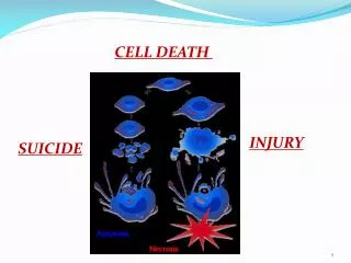
Apoptosis from Greek“apo” meaning “separation”&“ptosis” for “falling off” Kerr, J. F., et al.,(1972) Lecture V. Cell Birth & Death
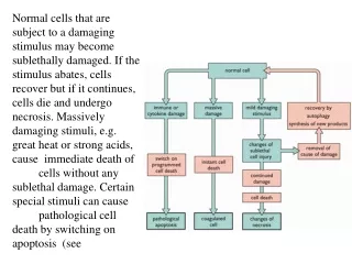
Types of Cell Death • Not Self-Initiated • Not Stereotypic • Can Be Slow • “Messy” • (injury can spread) Necrosis (Provoked) • Cell-Autonomous • Stereotypic • Rapid • “Clean” • (dead cells eaten) Apoptosis (Programmed) Ellis, R. E., et al., (1991) Lecture V. Cell Birth & Death
Removing a neuron’s targets, leads to its death Deprived Normal Hamburger, V. (1958,1977) Lecture V. Cell Birth & Death
Neuronal death is central for normal NS development Hamburger, (1975); Landmesser & Pilar, (1974); Cowan & Wenger, (1967) Lecture V. Cell Birth & Death
Neuron survival correlates with target innervation Not all neurons innervate targets Target Muscles Motor neurons Axon Outgrowth Development Progresses Target Innervation Neuronal Loss Lecture V. Cell Birth & Death
Target innervation determines which neurons survive Development Progresses More targets (more neurons) Fewer targets (fewer neurons) Lecture V. Cell Birth & Death
Mouse tumor (sarcoma) transplanted next to developing chick spinal cord causes axon sprouting consistent with a diffusible factor - a nerve growth factor Levi-Montalcini, R., & Hamburger, V. (1951) Lecture V. Cell Birth & Death
A quantitative functional assay for Nerve Growth Factor (NGF) activity, using explanted cultures of sensory ganglia Rita Levi-Montalcini Viktor Hamburger Stanley Cohen Levi-Montalcini, et al. (1954); Cohen, S., et al. (1954) Hollyday, M. (2001); Abbott, A. (2009). Cohen, S. (1987) Lecture V. Cell Birth & Death
NGF is the founding member of a large gene family of Neurotrophins (NTs), distantly related to insulin NGF binds as a dimer to its receptor Wiesmann, C., et al. (1999) Lecture V. Cell Birth & Death
NGF/Neurotrophins Signal through Trk (tyrosine kinase) Receptors Gene Activation/ Repression NGF/NT Trk Receptors (TrkA, TrkB, TrkC, p75) Apoptosis pathway Multiple Signaling Pathways Via kinases and scaffolding proteins (PIK3/AKT kinase) (PLC/PKC kinase) (Ras/MAP kinase) Intracellular Ca+2 release, modulation of ion channels Lecture V. Cell Birth & Death
C. elegans is the model organism for molecular genetic studies Bodywall Muscle Hypoderm Neurons Cuticular Cells Pharynx Intestine developmental time Vulva Gonad Germ Cells Muscle Neuronal Cell Death Lineages Sydney Brenner John Sulston H. Robert Horvitz Brenner, S. (1973) ; Sulston, J. E., & Horvitz, H. R. (1977); Sulston, J. E., et al. (1983); White, J. G., et al. (1982) Lecture V. Cell Birth & Death
Programmed Cell Death of single identified neurons can be followed in live worms P11aap Sulston, J. E., & Horvitz, H. R. (1977) Lecture V. Cell Birth & Death
(pro-survival genes + pro-apoptosis genes) (normal number of cells) (pro-survival genes + pro-apoptosis genes) (fewer cells) (pro-survival genes + pro-apoptosis genes) (extra cells) 2 Classes of C. elegans Cell Death Mutants WT Mutant class I Mutant class II Lecture V. Cell Birth & Death
ced-3(lf) reduced cell death (extra cells) viable ced-4(lf) reduced cell death (extra cells) viable ced-9(lf) ced-3(lf) reduced cell death (extra cells) viable ced-9(lf) ced-4(lf) reduced cell death (extra cells) viable Cell Death (pro-survival) gene (pro-apoptosis) genes ced-4 ced-9 ced-3 Genetic analysis of cell death genes in C. elegans defines a genetic pathway ced-9(lf) excessive cell death (fewer cells) animals die X as embryos X X X Ellis, R. E., et al., (1991); Ellis, R. E., & Horvitz, H. R. (1991) Lecture V. Cell Birth & Death
t(14;18) Chromosomal Translocation Causes Human B-Cell Leukemia by Overexpression of Bcl-2 Bcl-2 Chromosome 18 Ig Heavy Chain Chromosome 14 Bcl-2 Chromosome 18 Ig Heavy Chain Chromosome 14 Bcl-2 t(14;18) Chromosomal Translocation Stanley Korsmeyer Cleary, M. L., et al., (1986); Haldar, S., et al. (1990); Korsmeyer, S. J. (1992); Vaux, D. L., et al. (1988) Lecture V. Cell Birth & Death
The “core” Cell Death genes found in C. elegans are conserved as multigene families in vertebrates • Bcl-2: B-Cell Leukemia. • “Pro-survival” protein. • Inhibits release of • cytochrome C from • mitochondria (vertebrates). • Sequesters CED-4 • from cytoplasm (worms). ced-9 / Bcl-2: ced-4 / Apaf: • Apaf: Apoptosis activity factor. • “Adaptor” or “scaffold” protein. • Aggregates inactive procaspase, • causing auto-activation by proximity. • Requires cytochrome C, and ATP • for multimerization (vertebrates). ced-3 / Caspase: • Caspase: Cysteine • active-site, aspartate • cleavage-site, Protease. • “Terminator” protein. • Protease activity when activated by • proteolysis. Xue, D., & Horvitz, H. R. (1997) Yang, X., (1998) Xue, D., et al. (1996);Yuan, J. Y., & Horvitz, H. R. (1990) Lecture V. Cell Birth & Death
Molecular Model for Apoptosis mitochondria Bcl-2 (ced-9) single BH3 domain protein (egl-1) - - - (BH3 domains) Apaf (ced-4) Cytochrome C caspase (ced-3) (procaspase recruitment) - - (*Catalysis of the removal of auto-inhibitory caspase domain*) - - - - Apaf aggregation Inactive Procaspase recruited activated caspase (cascade) Death Lecture V. Cell Birth & Death
NGF is only one of multiple pathways to the “core” death mechanism, through many single-BH3 proteins Initiation of apoptosis by extracellular ligands (FAS, TNF) Single BH3 domain protein Single BH3 domain protein * Single BH3 domain protein * * * * “Core” apoptotic components NGF “Initiator” caspase-8 BCL-2 Gross, A., et al., (1999) Lecture V. Cell Birth & Death
Apaf/Cytochrome C Aggregate into a 7-Spoke Apoptosome Complex (“Wheel of Death”) Single-particle Electron Microscope Analysis WD-40 Apaf WD-40 CARD (caspase activation and recruitment domain) Cytochrome C Apaf gene +procaspase-9 (x7?) procaspase-9 Acehan, D., et al. (2002) Lecture V. Cell Birth & Death
“Pro-death” Single-BH3 domain proteins complex with Bcl-2 to release cytochrome C from mitochondria through “giant” mitochondrial ionic channels. BH3 pA Bcl-xL (Bcl-2 like) Diptheria Toxin (pore forming) Single-BH3 domain molecules integrate multiple signals that trigger apoptosis. Mitochondria integrate “Pro-survival” and “Pro-death”signals from a family of Bcl-2-like genes. (BH3) (BH3) Pro-survival Pro-death Schlesinger, P. H., et al., (2002), Fesik, (2000) Lecture V. Cell Birth & Death
Molecular Animation of Cell Death Mediated by the FAS pathway Berry, D. (2006) http://www.wehi.edu.au/education/wehi-tv/?page=2. Lecture V. Cell Birth & Death
What this Lecture was about • Programmed cell death (apoptosis) is a physiological mechanism distinct from necrotic cell death. • Apoptosis occurs widely during normal development of the nervous system. • Isolation of specific molecules involved in promoting growth and survival – “trophism,” e.g., Nerve Growth Factor (NGF). • What is the “death mechanism” that NGF (and other neruotrophins) inhibit? • Broader implications: controlled cell death in neuroembryology vs uncontrolled cell growth of cancer. • Gene homologies between organisms - humans and worms (nematodes) • Molecular models for apoptosis • How do trophic factors connect to this cell death pathway(s)? Lecture V. Cell Birth & Death
END Lecture V. Cell Birth & Death