Download
1 / 1
20 likes | 270 Views
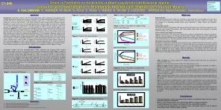
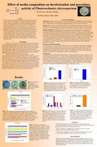
Effect of media composition on decolorization and peroxidase activity of Phanerochaetes chrysosporium. Erica Powless (Dr. Roy Ventullo) Wartburg College, Winter 2004. Azo dye. Abstract

E N D
Effect of media composition on decolorization and peroxidase activity of Phanerochaetes chrysosporium Erica Powless (Dr. Roy Ventullo) Wartburg College, Winter 2004 Azo dye Abstract Azo dyes are synthetic molecules containing an azo (-N=N-) functional group found in carpets, clothes, and plastics. The problem with these versatile, color-fast dyes is that they are allergenic, toxic, and may be carcinogenic, and they are not readily biodegradable in wastewater treatment. The lignin-modifying enzymes secreted by Phanerochaete chrysosporium (PC)are capable of decolorizing (degrading) azo dyes. The aim of this study was to determine the effect of media composition on decolorization and peroxidase activity. We hypothesized that decolorization would correlate with peroxidase activity since peroxidases are purported to be involved in decolorization. Decolorization of azo dyes was measured by growing PC in agar media of different composition. A simple medium consisting of corn syrup and yeast extract provided for the most rapid decolorization. A flourometric method was developed to measure peroxidase in the agar media. Peroxidase activity varied depending on the media used. This research adds to the understanding of how white-rot fungi degrade azo dyes and may contribute to the development of a treatment system for azo dyes waste. Materials and Methods Organism.Phanerochaete chrysosporium (PC), obtained from the lab of Dr. John Bumpus (University of Northern Iowa). Among its characteristics are the ability to carry out non-selective and non-stereospecific degradation of lignin, azo dyes, and pollutants to carbon dioxide at its high optimum temperature (40 C) for growth and activity. Azo Dyes. Three common azo dyes, Congo Red, Orange II, and Trypan Blue, representing the structural diversity of the dye group were used. Stock solutions of each azo dye were prepared by mixing 100 mg of the respective azo dye powder into 10 ml of sterile Nanopure® water. The azo dye powders were obtained as a gift from Dr. John Bumpus. Growth on solid media. Growth of PC and dye decolorization was followed using several media formulations. PC was grown on agar plates containing Malt Extract Agar (MEA), Potato Extract Agar (PEA), or Corn Syrup Yeast Extract Agar (CSYE). CSYE consists of 1g ml corn syrup, 10g ml yeast extract, 1.6g Bacto agar, and 100 ml Nanopure water. Media formulated by Pointing et al, (2002) and Martins et al. (2001), originally designed to be in the liquid form, were prepared as solid media by the addition of agar. Five mg of dye stock was added to 100 ml autoclaved media before the plates were poured. Plates were inoculated in the center of the plate with a PC suspension of spores obtained from a fully grown plate of PC. and incubated inverted in plastic covered boxes containing a water soaked paper towel (to create a high humidity environment) at 39 C. The growth of the fungus and decolorization of the dyes were recorded daily for two weeks. A scale of 0-4 was used to rank each of these processes, zero representing no growth or decolorization, one to three representing partial growth and decolorization, and four representing full growth or decolorization of the plates. Extraction of enzyme from agar plates. PC was grown for two weeks on each of the media. Peroxidase was extracted from the agar using the peroxidase kit reaction phosphate buffer. A sterile scalpel was used to cut a 25 mm x 20 mm plug from an agar plate exhibiting PC growth and decolorization. The plug of agar was then placed into a sterile glass tube (mortar) with two ml of 1X reaction buffer and ground with a motorized Teflon pestle. The homogenized solution was then transferred into two microfuge tubes and centrifuged at 6000 rpm for 3-4 minutes. The supernatant that resulted after centrifugation was then pipeted into another 1 mL centrifuge tube on ice for use in the enzyme assay. Peroxidase assay. The Amplex Red Hydrogen Peroxide/Peroxidase Assay Kit A-22188 (Molecular Probes, Eugene, Oregon) was used to measure the peroxidase activity of PC. A working solution of 100 uM Amplex Red reagent and 2.0 mM H2O2. served as the Amplex Reaction Mix. Reactions were run in Costar 96 well plates (Corning Incorporation, Corning, New York) and read in a Cary Eclipse Fluorescence Spectrophotometer (Varian Instruments, Walnut Creek, CA) equipped with a plate reader. Control wells contained 150 uL of 1X buffer and 50 uL of the Amplex Reagent Mix. Test wells typically contained 100ul buffered extract and 50 uL of Amplex Reagent Mix. The Amplex Reagent was added to start the reaction and plates were immediately placed in the plate reader (wavelengths: excitation 530nm, detection 590nm. Peroxidase activity (au/min) was determined from the slope of 20 min reaction curves. Introduction Phanerchaete chrysosporium (PC) is a ligninolytic fungus which produces the oxidative enzyme, lignin peroxidase. PC has been shown to degrade a wide variety of environmentally hazardous compounds, including nitroaromatics, polycyclic aromatic hyrdrocarbons, chlorinate organics, and azo dyes to carbon dioxide (Tatarko & Bumpus, 1998; Martins, Ferrerira, Santos, Queiroz, & Lima, 2001; Chagas & Durrant, 2001). Under nutrient limiting conditions, PC secretes lignin peroxidase that is capable of catalyzing the initial oxidation of several xenobiotics (Tuisel et al., 1990). Consequently, biodegradation of azo dyes by PC results in mineralization, but does not result in the formation of anilines as intermediates (Martins et al., 2001). Synthetic azo dyes are heavily used in the textile, paper, cosmetics, pharmaceutical and food industries (Chagas & Durrant, 2001). Azo dyes consist of one or more azo bonds (-N=N-) associated with one or more aromatic systems. Studies indicate that these dyes are toxic and also become harmful to the environment when they form carcinogenic and/or mutagenic aromatic amines (anilines) (Cripps, Bumpus, & Aust, 1990). Azo dyes are considered nondegradable under aerobic conditions with bacteria (Pasti-Grigsby, Pasczczynski, Goszczynski, Crawford, & Crawford, 1992). This makes it very difficult to remove azo dyes by conventional wastewater systems. The purpose of this study was to compare the decolorization of azo dyes by PC on various solid media and determine the activity of peroxidase released in the media. We hypothesized that the lignin-modifying peroxidase enzyme(s) secreted by PC would be more active in corn syrup yeast extract plates as faster decolorization occurs with this media. Results Figure 1: Decolorization of Trypan Blue incorporated into agar plates by PC. A, Control plate; B, CSYE medium; C MEA medium. Figure 5: This graphs shows the peroxidase activity of extracts from CSYE plates amended with Orange II. Control wells contained Nanopure water and Amplex (no enzyme). This data reinforced that we could measure activity . Figure 4: This graph compares peroxidase activity of extracts from CSYE media based on change in fluorescence (in arbitrary units, au) with time. The boiled extract showed little activity compared to the unamended and Trypan Blue amended CSYE. These results suggest that the addition of the dye to medium acts as an inducer of the peroxidase activity in PC. A C B Figure 7: A comparison of the peroxidase activity of extracts from unamended PEA plates. The addition of Trypan Blue (~500ug) to the reaction mix resulted in less apparent activity (green bar). This suggests that full decolorization of Trypan Blue and likely other dyes in the agar media is necessary to obtain an accurate measurement of peroxidase activity in plate extracts using the Amplex Red system. Figure 6: Peroxidase activity of extracts from plain and TB amended media. The high rate for PEA could be a result of the luxurious fungal growth on the plate. MEA, Martins, and Pointing showed little evidence of TB decolorization. At the time the assay was performed, the plates were not fully decolorized. We hypothesize that TB, as a substrate for the peroxidase, lowers the apparent peroxidase activity measured by the Amplex system. Figure 2: This graph shows the average time required for each medium to be completely covered with fungal hyphae (a 4 rating). Martins media amended with dye was the least effective medium for culturing PC whereas Pointing, PEA, MEA, and CSYE were usually within one to two days of each other for full growth coverage. Plates amended with Congo Red often took the longest to achieve full growth coverage, although MEA and PEA were covered in less than six days. Data was unavailable for the PEA Orange II plates due to contamination. The media formulated in our lab, CSYE, appears to be a good medium for growing PC. • Conclusions • CSYE overall appears to be a good medium for growing PC cultures and decolorizing Trypan Blue, Orange II, and Congo Red. The rapid decolorization allows for the detection of peroxidase with the Amplex system. • The low sensitivity of the Amplex Red Assay Kit does allow us to measure peroxidase activity in the different agar media tested. • The presence of dye appears to interfere with the Amplex Red reaction. Further work is needed to determine the extent of interference of residual dye. Figure 3: The average time required for complete decolorization (a 4 rating) of each agar medium [Legend is same as Figure 2].. Martins and Pointing consistently required more than 18 days for complete decolorization. This was also true for Congo Red amended MEA and PEA media. Due to the size and complexity of the structure of Congo Red, it was expected that complete decolorization would take longer than the other dyes. CSYE overall appeared the most efficient media for decolorizing Trypan Blue and Orange II and was marginally better with Congo Red (~15 days). Acknowledgements We would like to thank the Wartburg College Biology Department for the use of their supplies and equipment. A special thank you to Julie Paladino for ordering the necessary materials and equipment and John Bumpus at the University of Northern Iowa for gifts and guidance. We would also like to extend our gratitude to Warburg College for their funding through the Academic Partnership program and the R.J. McElroy Trust Program for their generous grant.