Schizophrenia
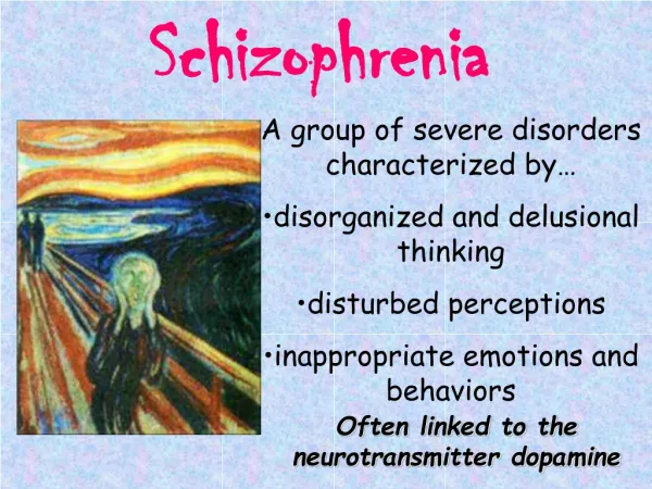
NIMH. Schizophrenia is a chronic, severe, and disabling brain disorder Affects 1.1% of the U.S. population age 18 and older in a given year. People with schizophrenia sometimes hear voices others don't hear, believe that others are broadcasting their thoughts to the world, or become convinced that


Schizophrenia
E N D
Presentation Transcript
1. Schizophrenia Stacy Zeigler
2. NIMH Schizophrenia is a chronic, severe, and disabling brain disorder
Affects 1.1% of the U.S. population age 18 and older in a given year.
People with schizophrenia sometimes hear voices others don�t hear, believe that others are broadcasting their thoughts to the world, or become convinced that others are plotting to harm them.
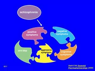
3. Symptoms develop in men- late teens or early twenties
women in the twenties and thirties, but in rare cases, can appear in childhood.
Hallucinations, delusions, disordered thinking, movement disorders, flat affect, social withdrawal, and cognitive deficits
4. Brain Research Reviews (2009) Superior temporal gyrus volume change in schizophrenia: a review on region of interest volumetric studies
5. Superior temporal gyrus (STG) Production, interpretation and self monitoring of language; implicated in AH
Superior temporal gyrus
1 of 3 gyri in temporal lobe
Brain volume/structure change may be linked to a brain region
6. Region of Interest (ROI) analysis STG structural differences
Advantages:
Anatomical validity, definition of landmarks in native space and quantitative measures of voxels
Limitations:
Labor intensive and time consuming
7. Studies considered Published up to July 2008 as an article
Compared schizophrenia patients with healthy group
Data on volume of STG and its subregions
Used ROI volumetric method
Individuals with schizophrenia and related diagnoses
Early onset schizophrenia included
Follow-up included
8. Details 2771 subjects
1444 patients
1327 controls
46 studies (5=follow up)
11.8 to 72 years old
Male patients 3 times more than females
Illness duration= 0.44 to 23.3 years
9. 24% of studies- no significant difference in STG volume and/or subregions between schizophrenic patients and controls
43% unileratal effects in STG or subregional volume change
Reduced on left side of STG more reported
37% bilateral reduction effect in STG or subregional volumes
6 studies- mixed effects (unilateral/bilateral)
12. Most showed reduced effect in STG or several subregional volumes
43% of studies- unilateral reduction
More pronounced on left side
Left STG- substrate of auditory and language processing and may be related to common symptoms
The review support STG or its subregions as candidate region related to hallucinations
13. Raij et al. (2009)-procedure 11 subjects with AVH and able to rate subjective reality
Practiced task then entered fMRI scanner
Cylinder shaped response keys in both hands
Each beginning and each end
If no AVH in 18 sec? rate the reality or loudness of latest AVH by moving cursor via response key
15. Analysis of coupling Tested coupling of IFG with other brain regions during AVH vs. non-AVH periods
One sample t test used to test the resulting contrast images for hallucination-related changes in the connectivity of IFG with other brain parts
Correlated contrast images with SRH across subjects
19. Strength of AVH-related activation in the IFG correlated with the SRH
Correlation of SRH with coupling between left IFG and left auditory cortex strongest in Heschl�s gyrus
Bilateral IFG signals correlated strongly with SRH
20. Brain (2008) 131: 3169-77 Auditory Verbal Hallucinations Predominantly Activate the Right Inferior Frontal Area
21. Method 24 Subjects
Frequent AVH and frequent moments without AVH
Right handed
Antipsychotic meds during study
17 males
7 females
18= schizophrenia
3= schizo-affective disorder
3= psychosis not otherwise specified
23. Method- continued Comprehensive Assessment of Symptoms and History (CASH)
Diagnosis
Edinburgh Handedness Inventory
The Positive and Negative Syndromes Scale (PANSS)
Symptom assessment
Psychotic Symptom Rating Scales- Auditory Hallucinations Rating Scale (PSYRATS-AHRS)
24. Procedure fMRI scans made continuously (8 min)
Patients squeeze balloon during AVH
Release when AVH subsided
Language activation measured (8 min)
Paced letter fluency task
Letter displayed on screen in front of them and patients silently generate word
Letters presented in 8 activation blocks
Each block= 30 sec
Each activation block- 10 different letters
1 letter every 3 sec
25. Procedure- continued 2 more letter fluency trials
Patients generate words aloud
Used to measure behavioural performance while they were in the scanner
Activation maps via Philips Achieva 3 Tesla Clinical MRI scanner
26. Procedure- continued 3D PRESTO SENSE sequence
Fast scan sequence
Full brain coverage in .609 sec
Combines 3D PRESTO pulse sequence with parallel imaging (SENSE) in 2 directions using a commercial 8 channel SENSE
SENSE= parallel imaging technique using multiple receiver head coils
800 3D PRESTO SENSE images aquired
27. Data Analysis Preprocessing
Reorientation and within-subject image realignment due to head motion
Comparing hallucinating and non-hallucinating periods
Squeezed balloon upon onset of hallucination
Stopped squeezing balloon when hallucinations stopped
28. Data Analysis- continued Letter fluency paradigm
Activation model created
Contrast activity when letter presented and rest periods
Following first level analyses, second level random- effects analyses conducted for both hallucination and letter fluency paradigm
Random effects group-wise conjunction analysis conducted
Identifies a �common processing component� by finding area activated in independent subtractions
29. Data Analysis- continued Lateralization indices calculated using individual t-tests
Lateralization indices= difference in �thresholded� signal intensity changes in L vs. R hemispheres divided by sum of �thresholded� signal intensity changes
Mask created using AAL (anatomical automatic labeling) atlas
Differences in indices compared via paired sample t-test
30. Data Analysis- continued Pearson�s correlations used to assess associations between:
Subjective loudness of AVH and activation of Heschl�s gyrus
Number of voices and activation of superior temporal gyrus
Lateralization index of AVH and degree to which emotional content of AVH was scored as negative
31. Results Subjects chronically psychotic
PANSS score average= 73
Average AVH several times/hour; lasting a few minutes
Hear voices inside and outside head (most)
Loudness- normal speaking
Most patients (18)- voices derogatory
6 patients- voices more neutral
32. PSYRATS-AHRS interview
33. During the scan- balloon task 18 hallucinations in 8 min
Duration- 20 sec
Total duration of hallucinations- 362 sec
34. Letter Fluency Task 96% correct performance
8 of the 24 patients- AVH during language and during resting blocks
35. fMRI Group analysis- multiple brain regions activated
Most extended activation in right inferior frontal area
Right insula and Broca�s homologue
Highly significant activation
Left motor cortex and right cerebellum
Significant activation during AVH
Left insula, bilateral supramarginal gyri, right superior temporal gyrus
Not significantly activated during AVH
Broca�s area and left superior temporal gyrus
38. Language Task Extensive activation of Broca�s area and contralateral homologue (lesser degree)
both extending into insula, bilateral temporal area (superior and middle gyri), left more than right, anterior cingulate gyri
Masks (created with AAL atlas) overlaid on group results
41. Group conjunction analysis Several areas activated
Right inferior frontal gyrus (including Broca�a homologue)
Right dorsolateral prefrontal cortex (DLPFC)
Left insula and right anterior insula
44. Lateralization Mean lateralization index
-0.11 for hallucination paradigm
0.14 for word generation task
Lower lateralization during AVH compared to word generation
Individual lateralization indices of hallucinatory activation not correlated to lateralization indices of word generation
46. Lateralization- continued No association with:
AVH loudness and Heschl�s gyrus activation
Number of voices and superior temporal gyrus
No difference in activation during AVH between individual with voices inside or outside head
More negative emotional content of voices associated with stronger lateralization of hallucinatory activation to right hemisphere
47. AVH AVH most extensive activation in right inferior frontal area (right insula and right homologue of Broca�s area)
Significant activation during AVH in superior temporal and supramarginal gyri (mostly right hemisphere), and left insula
Broca�s area or left superior temporal gyrus- no significant activation during AVH
48. Word production task Activitation of left inferior frontal area (Broca�s area and left dorsolateral prefrontal cortex)
Left insula, left superior and middle temporal gyri, anterior cingulate gyrus
Right side homologues activated, but to smaller degree
Activation during inner speech more extended compared to hallucinatory activity
Primarily results from difference in the applied paradigm
49. Corrections Number and duration of AVH differed
Variable and less extended activation
Conjuction analysis applied
50. AVH vs. language production AVH activate right homologues of language areas
Especially the insula and Broca�s area homologue
Normal language production activates frontal and temporal language areas in left hemisphere
Large inter-individual variability in lateralization of activity during AVH
Activation correlated with AVH negative emotional valence
51. Where do AVH come from? Previous reports- Broca�s area activation
AVH arise from speech production area
Right inferior frontal area associated with AVH
Left hemisphere dominates right in language production (right handed subjects)
Psychotic patients- AVH= single words or truncated sentences and negative emotions
AVH?right hemisphere language areas
May explain low linguistic complexity and derogatory content characteristic of AVH
52. Limitations Non-specific acoustic activation due to scanner sounds
Dampened activity in primary auditory cortex during AVH
Cerebral activation pattern due to AVH and motor activity
But for the right inferior frontal area to be activated due to motor activity would be unusual
53. http://www.youtube.com/watch?v=7zMz3l7IXKA
54. Other pics- sakai (review)
55. Robbins and Arnsten- review
57. Javitt (2009) review
58. Hugdahl et al (review)
60. Modinos et al.