Download
1 / 37
370 likes | 603 Views
The Muscular System (chapter 8). Specialized tissue that enable the body and its parts to move . Diagrams to know for this chapter…. Anterior View. Posterior View. Head and Face. TRIVIA……. How many muscles are there in the human body? Answer: 640 Muscles

E N D
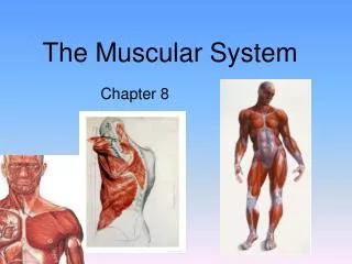
The Muscular System (chapter 8) Specialized tissue that enable the body and its parts to move.
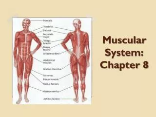
Diagrams to know for this chapter….. Anterior View Posterior View Head and Face
TRIVIA……. • How many muscles are there in the human body? • Answer: 640 Muscles • The muscles make up about 40 % of the body mass. • What is the longest muscle in the body? • Answer: The Sartorius • The Sartorius runs from the outside of the hip, down and across to the inside of the knee. It twists and pulls the thigh outwards.
TRIVIA……. • What is the smallest muscle in the body? • Answer: TheStapedius • The Stapedius is located deep in the ear. It is only 5mm long and thinner than cotton thread. It is involved in hearing. • What is the biggest muscle in the body? • Answer: The Gluteus Maximus • The Gluteus Maximus is located in the buttock. It pulls the leg backwards powerfully for walking and running.
Functions of the Muscles • Movement • Maintenance of posture and muscles tone • Propel body fluids and food • Generate heartbeat • Heat Production • Protect bones and internal organs
Muscle Classification • Functionally • Voluntarily – can be moved at will • Involuntarily – can’t be moved intentionally • Structurally • Striated – have stripes across the fiber • Smooth – no striations
3 types of muscles 2. cardiac 3. smooth 1. skeletal
Smooth Muscle • No striations • Single nuclei • Involuntary • Contracts slowly • Fibers are thin and spindle shaped • Found in circulatory, digestive, urinary, respiratory systems • Fatigue, but very slowly
Cardiac muscle • Striations • Each cell with central nuclei • Involuntary • Cells are branched and seem to be fused w/one another • Found ONLY in the heart • Healthy cardiac muscle NEVER fatigues…..or else.
Skeletal muscle • Fibers are long and cylindrical • Many nuclei • Striations • Voluntary • Attached to skeleton with tendons • Causes movement of bones at joints • Will fatigue (lactic acid)
Skeletal muscle functions 1. MAINTENANCE OF POSTURE AND MUSCLE TONE • Body position maintained because of contractions in our skeletal muscles. These contractions don’t produce movement yet hold our muscles in position. 2. HEAT PRODUCTION • contraction of muscles produces most of the heat required to maintain body temperature.
Skeletal muscle functions 3. MOVEMENT • Muscles move bone by pulling not pushing • Any movement is generally accomplished by more than one muscle • PRIME MOVER (AGONIST) = muscle responsible for a particular body movement. • SYNERGIST= A muscle that assists the action of the prime mover. • ANTAGONIST = A muscle that opposes a prime mover. (flex/extend)
Skeletal Muscle Structure • Composed of striated muscle cells (fibers) and connective tissue • Most muscles attach to two bones w/ a moveable joint in between • The attachment to the bone that does not move is the origin. • The attachment to the bone that moves is the insertion. • Tendons (dense fibrous connective tissue) anchor muscle to bone • Ligaments connect bone to bone at a joint • Bursae = sacs below tendons that secrete synovial fluid
Microscopic anatomy of skeletal muscle • Muscle cells (fibers) grouped in highly organized way. • Sarcolemma = membrane that surrounds the muscle cell • Muscle cells are filled w/2 threadlike proteins called myofilaments: • Myosin = thick • Actin = thin • These structures slide past one another causing the muscle cell to contract • Myofilaments are arranged in the cells in units called sacromeres.
Muscle contraction • Electrical impulses from the brain travel down a motor neuron. When it reaches the end, acetylcholine (chemical) is released. • Acetylcholine binds to special receptors on the muscle cell and cause and electrical impulse to spread over the cell. • The sarcomeres shorten and the muscle cell contract.
MUSCLE MYOFIBRIL MUSCLE FIBER SARCOMERE
Movement of muscles origin • Origin: the attachment of the muscle to the bone that remains stationary • Insertion: the attachment of the muscle to the bone that moves • Belly: the fleshy part of the muscle between the tendons of origin and/or insertion belly insertion
Categories of skeletal muscle actions CategoriesActions • Extensor Increases the angle at a joint • Flexor Decreases the angle at a joint • Abductor Moves limb away from midline of body • Adductor Moves limb toward midline of body • Levator Moves insertion upward • Depressor Moves insertion downward • Rotator Rotates a bone along its axis • Sphincter Constricts an opening
Practice these movements • Bend arm • - biceps contract • - triceps extend • 2. Straighten arm • - biceps extend • - triceps contract • 3. Bend knee • - quadriceps extend • - hamstrings contract
MORE MOVEMENTS 4. Straighten knee- quadriceps contract - hamstrings extend5. Crunches- abdomen contract - back muscles extend6. Point toes- calf muscle contract - shin muscle extend
Naming skeletal muscles • Location of the muscle • Shape of the muscle • Relative Size of the muscle • Direction/Orientation of the muscle fibers/cells • Number of Origins • Location of the Attachments • Action of the muscle
Muscles Named by Location • Epicranius (around cranium) • Tibialis anterior (front of tibia) tibialis anterior
Naming Skeletal Muscles Trapezius • Shape: • deltoid (triangle) • trapezius (trapezoid, 2 parallel sides) • serratus (saw-toothed) • rhomboideus (rhomboid, 4 parallel sides) • orbicularis and sphincters (circular) Deltoid Rhomboideus major Serratus anterior
Muscles Named by Size Psoas minor • maximus (largest) • minimis (smallest) • longus (longest) • brevis (short) • major (large) • minor (small) Psoas major
Muscles Named by Direction of Fibers • Rectus (straight) –parallel to long axis • Transverse • Oblique Rectus abdominis External oblique
Muscles Named for Number of Origins • Biceps (2) • Triceps (3) • Quadriceps (4) Biceps brachii
Muscles Named for Origin and Insertion Sternocleidomastoid originates from sternum and clavicle and inserts on mastoid process of temporal bone insertion origins
Muscles Named for Action • Flexor carpi radialis (extensor carpi radialis) – flexes wrist • Abductor pollicisbrevis (adductor pollicis) – flexes thumb • Abductor magnus – abducts thigh • Extensor digitorum – extends fingers Adductor magnus
Arrangement of Fascicles • Parallel • strap-like • ex: sartorius • Fusiform • spindle shaped • ex: biceps femoris
Arrangement of Fascicles • Pennate • "feather shaped” • Unipennate • ex: extensor digitorum longus • Bipennate • ex: rectus femoris • Multipennate • ex: deltoid
Arrangement of Fascicles • Convergent • ex: pectoralis major • Circular • sphincters • ex: orbicularis oris
There are about 60 muscles in the face. Smiling is easier than frowning. It takes 20 muscles to smile and over 40 to frown. Smile and make someone happy!!