The Muscular System
410 likes | 433 Views
This chapter explores the characteristics and functions of skeletal muscles, including their origin, microscopic anatomy, and contractions. Learn about the major muscles in the human body and the four functional groups of muscles. Understand how muscles produce movement, maintain posture, stabilize joints, and generate heat. Discover the interactions and roles of agonists, antagonists, synergists, and fixators in muscle activity. Dive into the detailed structure of skeletal muscles, including connective wrappings and microscopic anatomy. This comprehensive guide provides valuable insights into the intricate workings of the muscular system.

The Muscular System
E N D
Presentation Transcript
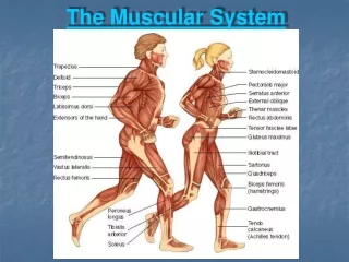
The Muscular System Textbook Reference: chapter 6
Unit ___ Goals Describe the characteristics and function of skeletal muscle Differentiate between a muscle’s origin and its insertion Identify the microscopic anatomy of skeletal muscle Differentiate between different muscular contractions Define the 4 functional groups of muscles Describe how a muscle is stimulated to contract and the mechanism of muscle shortening Identify the major muscles of the human body
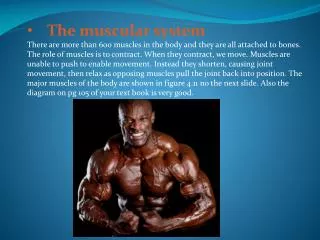
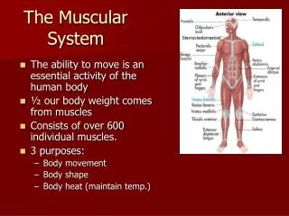
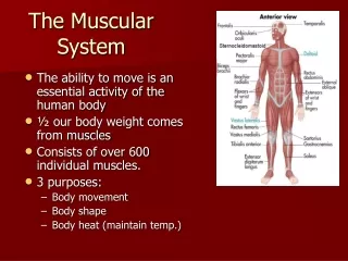
Muscles: the machines of the body • Skeletal Muscle Characteristics • Voluntary: move due to a conscious decision • Striated: striped in appearance • Multinucleate: Cells long; more nuclei • More mitochondria for energy production • 500+ muscles, 40-50% body weight
Skeletal Muscle Extras • Soft and fragile yet TOUGH • Exerts TREMENDOUS power • Power provided by bundles of muscle fibers wrapped in connective tissue • Connective tissue coverings allows for transmission of blood and nerves to muscles & provides support
Functional Characteristics of Skeletal Muscle • Produces movement: for locomotion, manipulation and responding to the external environment; also for expressing emotions • Maintaining posture: works continuously to fight downward pressure of gravity
Functional Characteristics of Skeletal Muscle • Stabilizing joints: muscle tendons stabilize and reinforce joints that have poor articulating surfaces • Generating heat: heat is a by-product of muscular activity; maintains normal body temperature
Skeletal Muscle Interactions • Muscles are arranged in the body to either work together or work in opposition of each other • Number 1 Rule of Muscle Activity: • MUSCLES ONLY PULL*** THEY NEVER PUSH! • In other words, muscle contractions are caused by SHORTENING of fibers
Insertion: the movable attachment of a muscle Examples: biceps tendon attachment to radial tuberosity Origin: the fixed, immovable point of attachment Examples: biceps tendon attachment to greater tubercle Skeletal Muscle Interactions A muscle contraction involves a muscle’s insertion moving toward its origin
4 Functional Groups of Skeletal Muscles • 1.) Agonists (prime movers): a muscle that provides the major force for providing a specific movement • Example: biceps brachii- prime mover of elbow flexion
4 Functional Groups of Skeletal Muscles • 2.) Antagonists: muscles that oppose/reverse a movement; relaxed when the agonist is active; helps regulate action of agonist by providing resistance • Can also be agonists • Example: the triceps brachii is the antagonist to the biceps during elbow flexion
4 Functional Groups of Skeletal Muscles • 3.) Synergists: “work together”; aids agonist by promoting the same movement or reducing unnecessary movements that might occur as the agonist contracts • Example: biceps brachii and brachioradialis in elbow flexion
4 Functional Groups of Skeletal Muscles • 4.) Fixators: a synergist that immobilizes a bone or a muscle’s origin • Example: erector spinae (muscles for posture) • All types of muscles work together to provide smooth, coordinated and precise movements. • Any one muscle can be in any functional group, dependant on its action
Detailed Skeletal Muscle • Deep fascia: sheet/band of dense connective tissue covering muscles • Epimysium: connective tissue covering many fascicles, all bound together
Fascicle: a bundle of fibers wrapped in perimysium Perimysium: connective tissue surrounding a bundle of muscle fibers Endomysium: connective tissue surrounding each muscle fiber Aponeuroses: sheetlike epimysia that connects muscle to each other and to cartilage/bone Detailed Skeletal Muscle
Detailed Skeletal Muscle • Tendon: cordlike band of epimysia connecting muscles to bones • Durable and able to cross bony projections • Small in diameter to fit in crowded joint spaces
Microscopic Anatomy of Skeletal Muscle • Muscle cell = muscle fiber • Sarcoplasm: cytoplasm of muscle fiber • Sarcolema: plasma membrane of muscle fiber • Sarcoplasmic reticulum: site of calcium storage used for contraction
Microscopic Anatomy of Skeletal Muscle • Sarcomere: contractile units of myofibrils, aligned end-to-end • Myofibril: contractile proteins of muscle cells; lie parallel along length of fiber • Thin myofilaments: actin, tropomyosin and troponin • Thick myofilaments: myosin
Microscopic Anatomy of Skeletal Muscle • Z line: separates sarcomeres; where actin filaments attach • I band:light bands; where the actin filaments are aligned; extends to tips of myosin filaments • A band: where the myosin filaments are aligned • H Zone: less dense portion of A band where thin filaments don’t overlap thick filaments
Nervous Stimulation of a Muscle Cell • Skeletal muscle cells possess unique abilities: • Excitability: can receive & respond to a stimulus • Contractility: they can shorten • Extensibility: lengthen or stretch • Elasticity: cells return to resting form after contracted or stretched • When a skeletal muscle contracts, it is told to do so by the nervous system
Nervous Stimulation of a Muscle Cell • Motor Neuron: nerve extending from spinal cord to muscle fiber • Motor Unit: one motor neuron and all the skeletal muscle cells it stimulates • Neuromuscular Junction: where the end of the nerve and the beginning of the muscle fiber meets • Synaptic cleft: space between nerve and muscle fiber • Motor end plate: highly folded sarcolema; lines synaptic cleft
Nervous Stimulation of a Muscle Cell • Neurotransmitter: a chemical that carries a signal from the nerve to the muscle; continues the nervous stimulation • Synaptic Vesicles: sacs at motor end plate that holds ACh • Acetylcholine (ACh): the neurotransmitter specific to muscles • Action Potential: an electrical current that can cause a muscular contraction
Nervous Stimulation of a Muscle Cell • The nervous system sends a neural signal to the motor unit. This causes a release of ACh into the neuromuscular junction, where it binds to receptors on the muscle cell. This causes an action potential to run throughout the muscle cell, initiating a muscle contraction.
Graded Responses of Skeletal Muscle Contraction • A graded response: the degree of muscle shortening dependant on • 1. The speed of muscle stimulation • 2. The number of muscle cells stimulated
Speed of Stimulation • Muscle Twitch: a single, brief contraction caused by a mistake of the nervous system • Summing of Contractions: when nervous impulses are delivered to a muscle at a rapid rate so it can’t relax between impulses; the contractions are summed(added) to create a smooth, strong contraction • Tetanus: no relaxation between contractions; smooth and sustained
Number of Cells Stimulated • Small, weak contractions: few cells initiated • Large, strong contractions: all motor units are active and all muscle cells stimulated
Types of Skeletal Muscle Contractions • Isotonic: same tone or tension • Myofilaments slide, muscle shortens & movement occurs
Types of Skeletal Muscle Contractions • Isometric: same length • Muscles DO NOT shorten • Myosin myofilaments do not move, tension in the fibers build • Muscle tries to move but cannot
Energy for Muscle Contractions • There are 3 different energy systems in the body that provide for muscle contractions • Definitions: • Creatine Phosphate (CP): high energy molecule only in muscles which help replenish energy stores • Adenosine Triphoshate (ATP): energy source in the body • Glucose: blood sugar • Glycogen: storage form of blood sugar
Energy for Muscle Contractions • Lactic Acid: a byproduct of an anaerobic pathway; causes muscle burning • Aerobic: using oxygen during activity • Anaerobic: not using oxygen during activity
Energy Systems • There are 3 ways energy is utilized for muscular activity: • 1. ATP-PC/Rapid Recovery • Anaerobic, lasting no more than 20 seconds • ATP production rate is rapid • ATP stores broken down rapidly and resupplied by creatine phosphate (transfers a phosphate to ADP) • More creatine stored in muscles than ATP
Energy Systems • 2. Lactic Acid/Anaerobic • Breaks down glucose for energy without using oxygen • Muscles are working too fast for oxygen & glucose to be delivered by aerobic respiration • Results in build up of lactic acid in muscles: promotes muscle fatigue and soreness
Lactic Acid/Anaerobic Continued • Muscle fatigue: when a muscle is unable to contract even though it is still being stimulated. • Oxygen Debt: occurs during prolonged muscle activity; can’t get oxygen fast enough for the muscle’s needs; lactic acid builds up leading to fatigue; repaid by breathing deeply until the muscles receive enough oxygen to get rid of lactic acid and make more ATP and creatine phosphate
Energy Systems • 3. Aerobic Respiration • During light exercise, ATP stores are regenerated by using oxygen • Can last for hours if glucose supplies are present • Used in endurance activities
Types of Skeletal Muscle Fibers • There are 2 different kinds: • 1. Slow twitch • For endurance • Speed of contraction is slow • Uses aerobic respiration • Low glycogen stores-more fats used • Slow rate of fatigue
Types of skeletal muscle fibers • 2. Fast twitch • Explosive movements for short distances • Speed of contraction very fast • Some aerobic, some anaerobic fibers • High glycogen stores but fatigues quickly The distribution of muscle fibers is not complete until the teenage years. It varies between muscles, within muscles and between people.