Download
1 / 49
490 likes | 517 Views
Learn about the three types of muscles - skeletal, cardiac, smooth - their structure, function, and mechanism of contraction. Dive into muscle structure and the molecular basis of muscle contraction. Discover how muscles work together and types of skeletal muscle fibers.

E N D
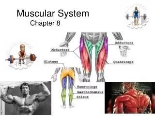
Muscular System Chapter 8
Learn your muscles Poke a Muscle
The 3 Muscle Types • The job of all muscles is to contract • They are all fibrous because cells are elongated • The 3 Muscle Types Are: • Skeletal Muscle • Cardiac Muscle • Smooth Muscle Overview Of Muscle
Skeletal Muscle • Cigar Shaped • Multinucleated • Striated • Voluntary • Can Be Involuntary When Reflexes Are Involved • Very Strong and Fast But Need Rest • Most Attached to Bone • Do not undergo mitosis. Once dead, dead.
Smooth Muscle Long thin nuclei and no striations
How are Muscles Structured? • Muscle Cells have a plasma membrane called a Sarcolemma. • The muscle fiber is enclosed in Endomysieum. (Endo= , Mys= ) • Many Muscle fibers bound together make a Fascicle. • The Fascicle is wrapped in a membrane called the Perimysieum (Peri= ). Pg. 174 Tortora
Muscle Structure Continued • Many fascicles are wrapped together by an Epimysium. (Epi= ) • Epimysia attach to tendons or Aponeuroses. (pg. 192: Tortora) • Tendons: Strong, Thin, and made up of collagen (dense connective tissue). Aponeuroses are sheet-like tendons.
Skeletal Muscle (pg. 174) Quiz Yourself
What is the major Organelle of the Muscle Cell? Pg. 176 • Myofibril(s)=Working unit of the muscle cell. Made of Subunits called sarcomeres. • Give muscles the striped or striated appearance • The light band is the I-band • The dark band is the A-band • Between the I-bands is the Z-line • Between the A-bands is the H-zone Match the Terms
Mechanism of muscle contraction • The above micrographs show that the sarcomere gets shorter when the muscle contracts • The light (I) bands become shorter • The dark bands (A) bands stay the same length
Take a long deep breath, its not that bad. • And remember a bicycle cannot stand alone, because it is two tired. • Now lets go on. • But lets first watch this short video clip. Overview of the Job of the Bands
So how do these bands work? • The myofibrils are surrounded by the sarcoplasmic reticulum, a specialized form of smooth endoplasmic reticulum that releases calcium. • They are made of bands of • Actin (the thin filaments) that make up the I-bands • Myosin (the thick filaments) that make up the A-bands
So what is the Molecular Basis of Muscle Contraction? (pg. 176) • 1) Nerve sends out Acetylcholine or ACh • 2) Motor Unit= All muscles triggered by nerve. (1 nerveTriggers 100’s of cells) • 3) The Sarcolema becomes permeable to Na+ • 4) Na+ causes an action potentialbecause it disturbs the electrical conditions of the sarcolema (pg.178)
How does ACh stimulate the muscle? • ACh causes the sarcolema to release Calcium (Ca+) • Ca+ binds to the actin causing it to change shape. • Myosine finds actin’s new shape attractive and grabs hold.
What happens after the Myosin grabs hold? (pg. 179) • Myosin’s head snaps towards the H-band of the sarcomere. • ATP releases and re-cocks the myosin • Only some myosin heads move at one time. • Over all: Pg. 181 Description of Muscle movement
How does the muscle relax? • When the action potential ends: • Sarcomere absorb Ca+ • ATP releases myosin heads • Actin takes on its former and less attractive shape. • Muscle Cells can relax Best Movie on Muscle Contraction Revisited Best Movie review of the muscle
Write one paragraph explaining how a muscle works. Do this from memory. This will help you learn. Paul Anderson Review
So, What is this Action Potential? • Action Potential • Electrical Current or Charge • In order to return the cell to its original condition, K+ is pumped into the cell by the sodium potassium pump. Sodium/ Potassium Pump
How do muscles work together? How do Muscles work Together? • Prime Mover: Major muscle doing the bulk of the work contracting. • Synergist: Group of muscles working together to contract. • Antagonist: Muscle that works against the prime mover and or synergists.
Summation and Tetanus • Twitch: a single action potential causing the muscle to contract for a millisecond and then relax. Latent...Contraction...Relaxation period • Summation effect: a second action potential arrives before the first is over. The contraction is larger. Wave summation • Tetanus: multiple summation effects causing a smooth sustained contraction. • Fatigue will set in and forces relaxation.
What is Recruitment? • Motor unit recruitment: various motor neurons in a whole muscle do not fire at the same time. Some are relaxed some are contracted. • Why? • Creates smooth muscle units not jerky movements. • Put your hand out and hold it still…..
Types of Skeletal Muscle Fibers • Slow…Slow Oxidative Fibers (SO) • Red, Make ATP Aerobiclatly, Fatigue resistant. Marathon runners getting out of a chair when old. • Fast • Fast oxidative –glycolytic Fibers (FOG). • Red, ATP by anaerobic Medium fast • Fast glycolytic Fibers (FG) • White. Anaerobic. Short, Fast, circuit trainers
Why can you crack your joints? Click here to find out!
Muscle Movements • http://www.healthchecksystems.com/exercise1.htm#hyper
Types of Muscle Movements Flexion: decrease in the angle between articulating bones. Extension: increase in the angle between articulating bones.
Abduction: movement of bone away from the midline Adduction: movement of bone towards from the midline
To exercise the: Quadriceps: straighten the knee
To exercise the: Hamstring Group Bend the knee. Flexes thigh. Includes the bicepts femoris
Exercise the: Trapezius : rotates shoulder., shoulder shrugs. Extends head.
Exercise for: Latissimus Dorsi: moves arm down and backwards.
Exercise for: Deltoid: abducts, flexes, extends & rotates arm at shoulder joint.
Exercise For: Gluteus Maximus extends and rotates thigh laterally at hip joint.
Exercises for: Oblique: Flexes vertebral column, contracts sides, rotates.
Exercises for: Rectus Abdominis: flexes vertebral column / compresses abdomen (think crunches)
Exercise for : Pectoralis Major Adducts and rotates arm medially. Extends arm at shoulder Push up: Works Pecs on The Up-Motion
Exercises for: Bicep contracts arm into L shape
Exercises for: Triceps …extends arm at shoulder and elbow
Now Quiz Yourself! See if you know your muscles… Another good Quiz site
Interesting Aspects: • Some of us may have a spare muscle: the Plantaris muscle 1 in 10 don’t. • Want to see it? Click the link. • Groin pull? Here is the problem: Plantaris In Action Groin Pull Or strain.


