
The Digestive System
The Digestive System. Digestive Processes. Ingestion – taking food into mouth. Propulsion – movement through alimentary canal (swallowing, peristalsis). Mechanical Digestion – Physical breakdown of food (chewing, churning).

The Digestive System
E N D
Presentation Transcript
Digestive Processes • Ingestion – taking food into mouth. • Propulsion – movement through alimentary canal (swallowing, peristalsis). • Mechanical Digestion – Physical breakdown of food (chewing, churning).
Chemical Digestion – Enzymatic breakdown of food (from complex to simple building blocks). • Absorption – transport of digested products from lumen of G.I. tract to blood and lymph (inside body). • Defecation – elimination of indigestable and waste products from body (feces).
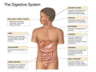
Salivary Glands Intrinsic (inside oral cavity) e.g., lips & cheeks. Extrinsic (outside oral cavity): “Mumps” 1)Parotid (largest) - a serous gland. 2)Submandibular - a serous gland. 3)Sublingual (smallest) - a mucus gland • Lingual amylase (breaks down starch). • Lysozyme – antibacterial agent in saliva.
Esophagus – Muscular tube, ~ 10 inches long. – Transition from skeletal to smooth muscle. (hence voluntary to involuntary) – Mucous glands in tela submucosa (layer) to lubricate bolus. – Outermost layer is Adventitia or Serosa. outside peritoneal cavity inside peritoneal cavity
Stomach – acidic (pH 2) storage of chyme. Mechanical Digestion continued (churning). Has 3 muscle layers, for churning. • Enzymatic Digestion of proteins occurs here (Pepsin breaksdown proteins). • Only Absorption of alcohol and aspirin. • Rugae allows for expansion when volume of contents increase.
Production and secretion of gastric juices controlled by CNS. e.g., Vagus nerve Parietal cells: make Hydrocholic acid (HCl) in gastric glands. Chief cells: make Pepsinogen, which is cleaved to pepsin (↑HCl), to digest proteins.
Small Intestine Duodenum Jejunum Ileum Increase Surface Area for Absorption 1) Plicae Circulares 2) Villi (Intestinal) 3) Microvilli • Lacteals absorption lipids Intestinal glands Goblet cells Stem cells
Villi of Small Intestine: Vascular Arcade (from superior mesenteric a.) Mesentery Proper - is a double layered serous membrane attached to the small intestine. • Roles: • - Supports branches of blood vessels. • - Supports lymphatics of the jejunum and ileum. • - Supports nerves of the jejunum and ileum.
The distinguishing features of each region of the small intestine.
The Large Intestine Begins as pouch inferior to end of ileum Ends at anus. Functions of Large Intestine: 1) Reabsorb water and compact feces. • 2) Absorb vitamins (helps make Vit K) electrolytes. • 3) Stores fecal matter. The Cecum: • Contains the Ileocecal valve and connected to appendix. The Colon: – Ascending, Transverse, Descending, Sigmoid.
The Colon • Lack of villi • Abundance of goblet cells • Abundance of mucous-secreting intestinal glands
1. Tunica Mucosa • A Mucous membrane • 1) Epithelium • 2) Lamina propria • 3) Muscularis mucosae 2. Tela Submucosa • Areolar Connective Tissue • Submucosal Plexus for Nervous innervation
3. Tunica Muscularis Externa • Smooth muscle layers • 1) Inner Circular Layer • 2) Outer Longitudinal Layer • Myenteric Plexus 4. Tunica Serosa (or Adventitia*) Serous membrane – aka visceral peritoneum • * Name depends on location: • Inside peritoneal cavity = serosa • Outside peritoneal cavity = adventitia
Tunica Mucosae • Lines digestive tract • Moistened by glandular secretions • Simple or stratified depending on area of tract • Pleated for expansion (Surface Area)
The Peritoneum: Two layers Visceral peritoneum (a.k.a. serosa) Parietal peritoneum - Lines inner surfaces of body wall Mesenteries: – Fused double sheets of peritoneal membrane to suspend portions of digestive tract: e.g. Greater omentum Lesser omentum Mesentery proper Transverse mesentery Sigmoid mesocolon
Retroperitoneal Structures – these are attached to posterior abdominal wall. e.g. • - Most of the Duodenum • - Ascending colon • - Descending colon • - Pancreas
Horizontal section through the upper abdomen showing the position of the liver relative to other visceral organs.
Muscularis Mucosae • Smooth muscle layer capable of plasticity • Ability to tolerate stretching • Visceral smooth muscle • Cells arranges in sheets • No motor innervation • Pace setter cells cause waves of contraction


