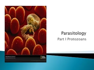
Parasitology
Parasitology. What is a parasite?. Definition: An animal which lives in (endoparasite) or on (ectoparasite) another animal (the host). Is almost always a different species from the host Depends on the host for food an causes some degree of injury. General characteristics.
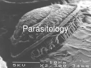

Parasitology
E N D
Presentation Transcript
What is a parasite? • Definition: • An animal which lives in (endoparasite) or on (ectoparasite) another animal (the host). • Is almost always a different species from the host • Depends on the host for food an causes some degree of injury
General characteristics • Tremendous reproductive capabilities • Have physical adaptations that enhance attachment to the host (i.e. suckers, hooks or clamps) • Possess various mechanisms for avoiding the host’s immune response • Exhibit complex life cycles often with multiple hosts
Typical Indirect parasite life cycle Primary Host Adult stage parasite infects host Infective larvae Able to infect primary host Egg/Spore Stage Transmission and spread into the environment Intermediate host Growth and developmental stage (may not cause damage to the host)
Negative affects on the host • Direct damage to host • inducing tissue and organ damage • Indirect effects • Stress causes an increase susceptibility to secondary infections • Direct damage can act as a portal for secondary infection • Parasite may serve as a carrier/vector for another viral/bacterial pathogen
Fish at surface gulping or piping Suggests parasites on gills Fish rolling/flashing suggests protozoan or worm infestation (internal or external) Lethargy or listlessness Suggests gill parasite Fish at bottom Suggests gill parasite, especially “Ich” Fin erosion/Lesions Indicative of external parasite Flared gills Indicative of gill parasite Excess mucus, fish “shimmies”/quivers, or is off feed General indication of disease Signs of Parasitic infections
Major Groups of Fish Parasites • Protozoa: single celled animals • Monogenetic Trematodes: Flukes (flatworms) with haptor (posterior attachement organ) an have simple life cycle (no intermediate host) • Digenetic Trematodes: Flukes (flatworms) with oral/ventral suckers and exhibit complex life cycles (involve intermediate hosts)
Major Groups of Fish Parasites • Cestodes (Tapeworms): worms with flattened/segmented bodies, head usually has suckers/hooks/suctional grooves • Nematodes (Roundworms): Thin elongated worms with cylindrical bodies covered by a rigid cuticle • Acanthocephala (Spiny-headed worms): bodies cylindical or fattened with anterior end bearing elaborate hooked proboscis
Major Groups of Fish Parasites • Copepods: crustaceans (sea lice) that may appear louse, worm, or grub like • Leeches: flattened or cylindrical, body segmented with anterior/posterior suckers • Glochidia: larval freshwater clams • Fungi: either as spores or as fungal hyphie
Flagellates Ichthyobodo (Costia) Ciliates Ichthyophthirius multifilis (“Ich”) Trichodinids Epistylis External Protozoa
External Protozoa • Common and usually occur in low numbers • Dense populations can cause serious epizootics (usually caused by some form of stress) • Symptoms include: -Irritation (flashing) -Erosion of scales -Erythema (reddening) -Hemorrhaging -Excess mucus production -white spots on skin • Control by chemical treatment
Myxobolus cerebralis (whirling disease) Ceratomyxa shasta Henneguya Internal Protozoa
Infects cultured and wild salmonids Specific tropism for cartilage Infection can result in axial skeleton and neural damage Myxobolus cerebralis
Myxosporea Found in marine and freshwater environments Only infects salmonids Susceptibility varies Clinical signs vary among infected salmonid species Identified by spore size, shape, and location Ceratomyxa shasta
Myxosporea Ovoid, spherical, or lenticular spores Usually cysts form around spores Henneguya
General Myxosporidean lifecycles • landmark discovery by Wolf & Markiw in 1984 • a fish myxosporean alternates with an actinosporean from an oligochaete worm • both spore types represent alternate lifecycle stages of the one organism • morphologically distinct spores are genetically identical
General Diagnostic Procedure • Presumptive ID: • Wet preparation • Histology • Site of infection • Spore morphology • Confirmation of ID: • Molecular methods • Any level of infection, all stages, definitive
Monogenetic Gyrodactylus sp. Digenetic Bolbophorus damnificus is often referred to as the “catfish trematode” Misnomer because there are about 30 trematodes found in the channel catfish and because it is also found in the fathead minnow
Digenetic Trematodes • Adult flukes reside in fish, birds, or mammals • Flukes lay eggs that pass through the definitive host, eggs hatch to a ciliated miracidia • The miracidia will develop to a cercariae if in contact with a snail or mussel • If the cercariae contacts invertabrate of fish hose it will encyst as a metacercaria
Cestodes • GI tract of fish, bird or Mammal • Eggs are laid to water and are eaten or hatch into a coracidium (C) and are then eaten by an invertabrate host • Larval development to a proceroid or a pleroceroid occurs invertabrate • Final host becomes infected by ingesting invertebrate
A.colex of Bothriocephalus acheilognathii from carp, Transvaal, South Africa (by courtesy of J.G. Van As). B. B. acheilognathii, whole worm (living) from farmed carp, Israel. C. Embryonated eggs of b. D. Ligula sp. from Rastrineobola argenteus from L. Victoria. Infected fish are recognized by their inflated abdomen (top fish) and may accommodate even three worms (bottom group).
Nematodes C. philippinensis egg C. philippinensis adult
Unembryonated eggs are passed in the stool (1) and become embryonated in the environment (2); after ingestion by freshwater fish, larvae hatch, penetrate the intestine, and migrate to the tissues (3). Ingestion of raw or undercooked fish results in infection (4). The adults reside in the human small intestine mucosa (5). The females deposit unembryonated eggs (can become embryonated) (6). Also infects fish eating birds (7). Capillaria philippinensis
Acanthocephala • A) GI tract of Fish, Acanthor larva released • B) Eaten by invertabrates and produces a cystacanth (C) • If eaten by suitable host, the cystacanth will develop into an adult
Neoechinorhynchus rutili Adult female Adult male
Leeches (Hirudinea) Leech with brood attached Top view
Leech Characteristics • Primarily occur in freshwater • Most are predators or scavengers which feed on fluids or soft tissues of live or dead invertebrates • Generally have 34 body segments and an anterior and posterior sucker • Parasitic leeches attach temporarily • Cause little noticeable harm
Hemorrhaging Inflammation Edema Ulceration Fibrosis Hyperplasia Necrosis Irritation Weight loss Some can be vectors of other parasistes Hirudinea Problems
Copepods Lernaea sp. Salmonicola sp.
Copepods are a subclass of Crustaceans • Sexes are usually separate – with sexual dimorphism present • Heavy infections can cause severe damage to skin, muscle, and gill tissues • Can also lead to secondary infections, anemia, emaciation, and mortality
Copepoda • Mature copepods release eggs (B) that hatch to larvae (C) • D) After molting a copepod stage is formed and may attach to a host (E)
Glochidia • Larvae attach to gills or skin • Live as parasites then drop off and live independently • Some modify mantle tissue to help find host
Glochidia Lampsilis reeveiana Glochidia attaching to gill tissue
Fungi • Saprolegnia “water molds” • Worldwide in freshwater • Appear as whitish cottony-like growths • Considered secondary invaders • Can attach to eggs and fish • Can be controlled with chemicals
Aphanomyces invadans Hyphae Germination Cyst Sporangia Sporulation 2° Zoospore 1° Zoospore 1º cyst