Download

1 / 46
470 likes | 512 Views
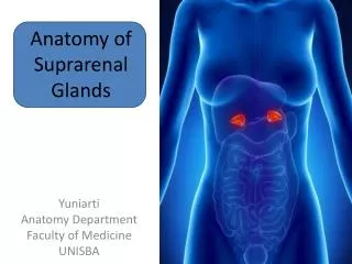
Understanding the structure and function of the suprarenal (adrenal) glands, which secrete steroid hormones and catecholamines. Explore their location, blood supply, secretory regions, and nerve innervation.

E N D
Gross Anatomy of the suprarenal glands Dr. shatarat. The University of Jordan
secrete both steroid hormones and catecholamines • Suprarenal (adrenal) Glands • ADRENAL GLANDS Dr. shatarat. The University of Jordan
The adrenal glands are two small triangular structures located Retroperitoneally at the upper poles of the kidneys. Dr. shatarat. The University of Jordan
The adrenal glands are covered with a thick connective tissue capsule from which trabeculae extend into the parenchyma carrying blood vessels and nerves. Dr. shatarat. The University of Jordan
They are found on the posterior parietal wall, on each side of the vertebral column, at the level of the 11ththoracic rib And lateral to the first lumbar vertebra • They have a flattened triangular • shape and are embedded in the perirenal fat at the superior poles of the kidneys. • lie immediately superior and slightly anterior to the upper pole of the kidneys • The suprarenal glands each weigh approximately 5 g (the medulla contributes about one-tenth of the total weight). Dr. shatarat. The University of Jordan
The secretory parenchymal tissue is organized into two distinct regions The cortex is the steroid-secreting portion. It lies beneath the capsule and constitutes nearly 90% of the gland by weight The medulla is the catecholamine-secreting portion. It lies deep to the cortex and forms the center of the gland. Dr. shatarat. The University of Jordan
Blood supply Dr. shatarat. The University of Jordan
Blood supply of the adrenals • Each gland receives 3 arteries • Superior suprarenal a. from the inferior phrenic artery • Middle suprarenal a. • from the abdominal aorta. • Inferior suprarenal a. • from the renal artery. • The suprarenal gland receives the highest blood supply in the body/gm of tissue. Dr. shatarat. The University of Jordan
The capsule is penetrated by ~ 60 arterioles. short capsular capillaries that supply the capsule. The superior, middle, and inferior suprarenal arteries In the capsule they branch forming a system that consists of B)intermediate fenestrated cortical sinusoidal capillaries that supply the cortex C) long medullary arterioles that traverse the cortex traveling within the trabeculae, and bring arterial blood to the medullary capillary sinusoids. Dr. shatarat. The University of Jordan
The medulla thus has a dual blood supply arterial blood from the medullary arterioles and “venous” blood from the cortical sinusoidal capillaries that have already supplied the cortex. Dr. shatarat. The University of Jordan
Arterial and venous capillaries within the adrenal gland help to integrate the function of the cortex and medulla. For example, cortisol-enriched blood flows from the cortex to the medulla, where cortisol enhances the activity of phenylethanolamine-Nmethyltransferasethat converts norepinephrine to epinephrine. Extra-adrenal chromaffin tissues lack these high levels of cortisol and produce norepinephrine almost exclusively The largest cluster of chromaffin cells outside the adrenal medulla is near the level of the inferior mesenteric artery and is referred to as the organ of Zuckerkandl, which is quite prominent in fetuses and is a major source of catecholamines in the fi rst year of life An example of extra-adrenal chromaffin tissues Dr. shatarat. The University of Jordan
Venous drainage of the adrenal glands is achieved via the suprarenal veins: The venules that arise from the cortical and medullary sinusoids drain into the small adrenomedullary collecting veins that join to form The Large Central Adrenomedullary Vein which then drains directly into : Dr. shatarat. The University of Jordan
The right suprarenal vein (short) drains into the inferior vena cava Why? Dr. shatarat. The University of Jordan
Rt. • The left suprarenal vein (longer) drains into the left renal vein or the left inferior phrenic vein. Lt. Why? Dr. shatarat. The University of Jordan
only one suprarenal vein exists for each adrenal gland Dr. shatarat. The University of Jordan
Read only Normal variations in the adrenal gland A) arterial supply via three arteries b) arterial supply without tributary from the A. ranalis c ) arterial supply without a direct branch of the Aorta Dr. shatarat. The University of Jordan
Nerve supply Dr. shatarat. The University of Jordan
Relative to their size, the adrenal glands have a richer innervation than other viscera 1-The sympathetic preganglionicfibers for these glands are the axons of cells located in the intermediolateralcolumns of the lowest two or three thoracic and highest one or two lumbar segments of the spinal cord 2-They emerge in the anterior rootlets of the corresponding spinal nerves; pass in the white rami communicantesto the homolateral sympathetic trunks; and leave them in The greater splanchnic nerve lessersplanchnic nerves least thoracic splanchnic nerves first lumbar splanchnic nerves Dr. shatarat. The University of Jordan
3-They run to the celiac aorticorenal renal ganglia 4-Some fibers end in these ganglia, but most pass through them without relaying The preganglionic sympathetic fibers end directly around the medullary cells because these cells are derived from the sympathetic anlage and are the homologues of sympathetic ganglion cells. Other fibers innervate the adrenal vessels, including the central vein. Dr. shatarat. The University of Jordan
Catecholamines are released from the adrenal medullary and sympathoneuronal systems—both are key components of the fight-or-flight reaction This reaction is triggered by neural signals from several sites in the brain (e.g., the hypothalamus, pons, and medulla), leading to synapses on cell bodies in the intermediolateral cell columns of the thoracolumbar spinal cord. The preganglionic sympathetic nerves leave the spinal cord and synapse in paravertebral and preaortic ganglia of the sympathetic chain. Preganglionic axons from the lower thoracic and lumbar ganglia innervate the adrenal medulla via the splanchnic nerve ACETYLCHOLINE is the neurotransmitter in the ganglia, and the postganglionic fiber releases NOREPINEPHRINE. The chromaffin cell of the adrenal medulla is a “postganglionic fiber equivalent,” and its chemical transmitters are epinephrine and norepinephrine. Dr. shatarat. The University of Jordan
Parasympathetic fi bers are conveyed to the celiac plexus in the celiac branch of the posterior vagal trunk, and some of these are involved with adrenal innervation and may relay in ganglia in or near the gland Dr. shatarat. The University of Jordan
Embryology 1-Development of the cortex of the suprarenal gland Dr. shatarat. The University of Jordan
Two origins Embryologically, the cortical cells originate from mesodermalmesenchyme, whereas the medulla originates fromectodermal origin (neural crest cells) that migrate into the developing gland Dr. shatarat. The University of Jordan
Week 4 – 6 : start from mesoderm adjacent to urogenital ridge This origin (near to urogenital ridge) accounts for the presence of accessory para-testicular and para-ovarian accessory cortical masses Explain the presence of accessory para-testicular and para-ovarian accessory cortical masses? Dr. shatarat. The University of Jordan
At the beginning of 8th week of development mesothelial cells proliferate and differentiate into large acidophilic cells which surround the medullaryprimordium and form the fetal or primitive suprarenal cortex • Embryology • Definite cortex develop into functional adrenal cortex Dr. shatarat. The University of Jordan
At the end of the 3ed month of development a second wave of smaller basophlicmesothelial cells surround the original acidophilic cell mass. These smaller cells form the definitive cortex of the gland The small basophilic cells will form the future glomerular and fascicular zones of the definitive cortex • Fetal cortex produce steroid during gestation After birth, the fetal cortex regresses rapidly, except for its outer layer which differentiates into the reticular zone of the cortex Dr. shatarat. The University of Jordan
Prior to month 5 of intrauterine development The cortex appears to develop autonomously After the 5th month, the development of the adrenal gland development depends on hypophysealcorticotropic hormone (ACTH) Therefore, In case of anencephaly Anencephaly: is a serious birth defect in which a baby is born without parts of the brain and skull. It is a type of neural tube defect (NTD). As the neural tube forms and closes, it helps form the baby’s brain and skull (upper part of the neural tube), spinal cord, and back bones (lower part of the neural tube) Read only has little effect before month 5 of fetal life since development of the adrenal up to this point appears to be autonomous After month 5, development of the fetal cortex cannot occur without ACTH, thus, in the anencephalic, there is an involution of the adrenal cortex leading to agenesis or hypoplasia note agenesis : refers to the failure of an organ to develop during embryonic growth Dr. shatarat. The University of Jordan
IN HYDROCEPHALUS s a condition in which there is an accumulation of cerebrospinal fluid (CSF) within the brain. The hypothalamus is undamaged. The adrenals develop normally Dr. shatarat. The University of Jordan
The origin of the cortex of the suprarenal gland is (near to urogenital ridge) which explains the presence of accessory para-testicular and para-ovarian accessory cortical masses True accessory adrenal glands, consisting of both cortex and medulla, are rarely found in adults. When they are present, they may be within the celiac plexus or embedded in the cortex of the kidney.. Adrenal rests, composed of only cortical tissue, termed cortical bodies, occur frequently and are usually located near the adrenal glands. In adults, accessory separate cortical or medullary tissue may be present in the spleen, in the retroperitoneal area below the kidneys, along the aorta, or in the pelvis. Dr. shatarat. The University of Jordan
Because the adrenal glands are situated close to the gonads during their early development accessory tissue may also be present in the attached to the ovary, or in the broad ligament of the uterus. Although one adrenal gland may be absent occasionally, complete absence of the adrenal glands is extremely rare spermatic cord, attached to the testis in the scrotum Dr. shatarat. The University of Jordan
Embryology 2-Development of the MEDULLA of the suprarenal gland Dr. shatarat. The University of Jordan
Ectodermal cells arise from the neural crest and migrate from their source of origin to differentiate into sympathetic neurons of the autonomic nervous system. However, not all of the cells of the primitive autonomic ganglia differentiate into neurons. Some become endocrine cells, designated as chromaffin cells because they stain brown with chromium salts Certain chromaffin cells migrate from the primitive autonomic ganglia adjacent to the developing cortex to give rise eventually to the medulla of the adrenal glands. When the cortex of the adrenal gland has become a prominent structure (during the seventh week of embryogenesis), masses of these migrating chromaffin cells come into contact with the cortex and begin to invade it on its medial side. By the middle of fetal life, some of the chromaffi n cells have migrated to the central position within the cortex Dr. shatarat. The University of Jordan
Some chromaffin cells also migrate to form paraganglia, collections of chromaffin cells on both sides of the aorta. The largest cluster of chromaffin cells outside the adrenal medulla is near the level of the inferior mesenteric artery and is referred to as the organ of Zuckerkandl, which is quite prominent in fetuses and is a major source of catecholamines in the fi rst year of life Dr. shatarat. The University of Jordan
Congenital anomalies of the suprarenal gland Dr. shatarat. The University of Jordan
Fusion of the suprarenal glands: seen when kidneys are also fused across the midline Dr. shatarat. The University of Jordan
Congenital adrenal hypoplasia usually manifests itself shortly after birth with many of the symptoms of Addison's disease Agenesis of the adrenal: unilateral agenesis of the gland is almost always associated with agenesis of the kidney on the same side Dr. shatarat. The University of Jordan
Imaging of the suprarenal gland Dr. shatarat. The University of Jordan
The adrenal gland is the fourth most common site of metastasis, and adrenal metastases may be found in as many as 25% of patients with known primary lesions Adrenal cortical adenoma can be diagnosed with a high degree of accuracy: the specificity of imaging studies ranges from 95-99%, and the sensitivity is greater than 90% http://emedicine.medscape.com/article/376240-overview Unenhanced CT scan through the level of the adrenal glands shows normal appearing bilateral adrenal glands in the suprarenal fossa. The glands take on the appearance of an upside down "V" or "Y" often (arrows). Dr. shatarat. The University of Jordan
Histology Dr. shatarat. The University of Jordan