Download
1 / 23
290 likes | 542 Views
Explore the anatomy and physiology of the salivary glands, including the major glands like Parotid, Submandibular, and Sublingual, their embryological development, histology, and function in saliva production. Learn about their innervation and arterial supply.

E N D
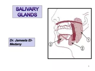
3 major glands: • • Parotid • • Submandibular • • Sublingual • • Plus many accessory glands in mucosa • • Exocrine glands that secrete directly into the oral cavity • •Important for lubricating mouth, remineralizing teeth, breaking down food • • Innervated by sensory, parasympathetic, and sympathetic fibres
Embryology • 6th to 8th week of gestation • Begin as oral ectodermal outpouchings into mesenchyma – point of origin is duct orifice • Parotid – first to develop & last to become encapsulated
Anatomy - Parotid • Largest salivary gland • Arbitrarily separated into two “lobes” by facial nerve • Compartment • Superior - Zygoma • Posterior – EAC, tail extends over SCM/mastoid • Inferior - Styloid process and muscles, carotid sheath • Anterior – Masseter and buccal fat pad
Anatomy - Parotid • Parotid fascia – continuation of superficial layer of deep cervical fascia • Superficial – spreads from masseter to SCM to zygoma • Deep – inferiorly forms stylomandibular membrane and connects styloid posteriorly to mandible anteriorly • Inelastic fascia, sends septations into gland preventing easy dissection
Anatomy - Parotid • Stensen duct – arises from anterior gland, 1.5 cm below zygoma • 4 – 6 cm long • Runs over masseter • Pierces buccinator at ant. border of masseter • Orifice is at level of 2nd maxillary molar • Buccal branch of VII parallels duct
Anatomy - Parotid • Arterial Supply – transverse facial artery (branch of superficial temporal) • Venous – retromandibular vein • Lymphatic: • Superficial layer below capsule – drains parotid, scalp, eyelids, EAC • Deep layer within gland – drains parotid, NP, middle ear, soft palate
Anatomy - Parotid Localization of CN VII • Tragal pointer • Tympanomastoid suture • Posterior belly Digastric • Styloid process • Retrograde dissection
Anatomy - Parotid Histology of the Parotid Gland • compound racemose gland • large number of ducts arranged in branching system • Mostly serous alveoli with cuboidal- or columnar-lined ducts
Important Points about the Parotid • Largest of the salivary glands • Mostly watery serous secretion • Closely associated with facial nerve and vasculature of the face • Parotid duct opens through cheek above upper second molar • Produces most stimulated saliva
Anatomy - Submandibular • Second largest salivary gland • Submandibular triangle: • inferior mandible • ant/post bellies of digastric • wraps around myelohyoid to form superficial and deep lobes
Anatomy - Submandibular • Wharton duct; • From medial surface – courses between myelohyoid and hyoglossus to FOM • Above CN XII / Below CN V3 (lingual) • 5 cm long • Orifice is lateral to lingual frenulum
Anatomy - Submandibular • Arterial Supply – Facial artery • Venous Drainage – Anterior facial vein • Lymphatics: • around gland in fascia • drain to deep cervical chain
Anatomy - Submandibular • Histology of the submandibular gland • Mixed gland with • serous and mucous alveoli • Ducts and lobules present, but not as extensive as in parotid • Demilunes are granular serous secreting cells
Anatomy – Sublingual • Sublingual gland • In FOM - above myelohyoid and between mandible and geniglossus • No fascia • Drains via 10 ducts of Rivinus into FOM along the sublingual plica • Arterial – Lingual/Facial • Venous – Lingual/Facial • Lymphatics – Submandibular nodes
Anatomy - Minor Salivary • Minor Salivary Glands • 600 – 1,000 lining entire oral cavity, BOT, tonsillar pillars and tonsils (Weber glands) • Each gland has own simple duct • Blood supply and venous drainage by location
Saliva • Acts as lubricant, wetting agent, prevents irritation and protects dentition • Isotonic fluid created by acini – becomes hypotonic as moves down duct (higher flow rates time for modification) • Active secretion of IgA, lysozyme, salivary peroxidase – innate immunity • Salivary peroxidase – glycolysis • Mucin – mucus provides moisturization of mucosa • Actively secreted by cell hyperpolarization
Saliva • Parotid: Serous cells only – Watery saliva • Submandibular: Mucinous and serous cells – Viscous saliva • Minor Salivary: Mixture serous and mucinous • Sublingual: Mucinous only • Salivary flow: • Unstimulated: • 69% submandibular, 26% parotid, 5% minor • rate - 0.001 – 0.2 ml/min • Stimulated: • Production reverses – 2/3 from parotid • Increases to 0.18 to 1.7 ml/min • Averages 1,000 – 1,500 ml/day
Function of Saliva • Moistens oral mucosa • Moistens & cools food • Medium for dissolved food • Buffer (HCO3) • Salivary peroxidase - Glycolysis • Digestion (Amylase, Lipase) • Antibacterial lysozyme, IgA,Peroxidase, FLOW) • Mineralization • Protective for dentition
Innervation of the Salivary Glands Main control by parasympathetic system: Submandibular and sublingual: Via chordatympani of facial nerve through submandibular ganglion Parotid: Glossopharyngeal nerve via otic ganglion Sympathetic fibres through cervical ganglion then follow blood vessels to glands
Factors Affecting Salivary Flow Important Factors Unimportant Factors • Degree of hydration • Body position • Exposure to light • Olfaction • Previous stimulation • Circadian rhythms • Drugs • Gender • Age (above 15 years) • Body weight • Gland size • Psychic effects • --thought/site of food • --appetite • --mental stress • Functional stimulation