BILIARY PASSAGES
BILIARY PASSAGES. Objectives: 1. The student should be able to identify & describe the histological features of intrahepatic biliary passages . 2. The student should be able to identify & describe the histological features of extrahepatic bile ducts .

BILIARY PASSAGES
E N D
Presentation Transcript
BILIARY PASSAGES Objectives: 1. The student should be able to identify & describe the histological features of intrahepaticbiliary passages. 2. The student should be able to identify & describe the histological features of extrahepatic bile ducts. 3. The student should be able to identify & describe the histological structure of gall bladder.
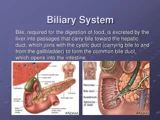
Biliary Passages Intrahepatic passages: 1- Bile canaliculi. 2- Bile ductules (canals of Hering). 3- Interlobular bile ducts. Extrahepatic passages: 4- Right & left Hepatic ducts. 5- Common hepatic duct. 6- Common bile duct.
Bile Canaliculi • Narrow channels located between hepatocytes, limited only by the cell membranes of 2 hepatocytes. • They are the first portions of the bile duct system. • Microvilli project from the hepatocyte into the bile canaliculi, thus increasing the surface area. • Tight junctions between the cell membranes of the 2 hepatocytes prevent leakage of bile.
Bile Ductules (Canals of Hering) • Near the peripheral portal areas, bile canaliculi empty into bile ductules composed of cuboidal epithelial cells called cholangiocytes. • After a short distance, these ductules collect and end in the interlobularbile ductsin the portal areas.
Interlobular Bile Ducts • Are in the portal areas. • Lined by simple cuboidal epithelium (becomes simple columnar epithelium near the portahepatis). • Interlobular bile ducts merge to form larger ducts, which eventually unite to form the right and left hepatic ducts.
Common Hepatic Duct • Formed by union of the right & left hepatic ducts. It joins the cystic duct, arising from the gallbladder, forming the common bile duct. • Similar in structure to the wall of gall bladder and other extrahepatic bile ducts. • Mucosa: • Epithelium: Simple columnar. • Lamina propria. • Muscularis:bundles of smooth muscle fibers in all directions. • Adventitia.
GALL BLADDER A saclike structure that stores, concentrates and releases bile. Its wall is formed of: • Mucosa: highly folded. • Simple columnar epithelium. • Lamina propria: contains mucous glands in the neck of gall bladder. • Muscularis: bundles of smooth muscle fibers oriented in all directions. • Serosa or adventitia.