LIVER AND BILIARY PASSAGES
540 likes | 842 Views
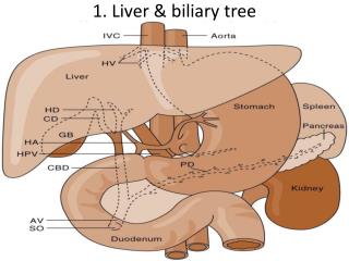
LIVER AND BILIARY PASSAGES. Dr. Mujahid Khan. Liver. Liver is the largest gland in the body Its basic functions are production and secretion of bile Metabolic activities related to carbohydrate, fat, and protein metabolism Filtration of the blood

LIVER AND BILIARY PASSAGES
E N D
Presentation Transcript
LIVER AND BILIARY PASSAGES Dr. Mujahid Khan
Liver • Liver is the largest gland in the body • Its basic functions are production and secretion of bile • Metabolic activities related to carbohydrate, fat, and protein metabolism • Filtration of the blood • Remove bacteria and other foreign particles that have gained entrance to the blood from the lumen of the intestine
Liver • The liver synthesizes heparin, an anticoagulant substance, and has an important detoxicating function • It produces bile pigments from the hemoglobin of worn-out red blood corpuscles • Secretes bile salts which are conveyed to the duodenum by the biliary ducts
Location • It is soft and elastic, occupies the upper part of the abdominal cavity just beneath the diaphragm • Its greater part is situated under cover of the right costal margin, and the right hemidiaphragm separates it from the pleura, lungs, pericardium, and heart • It extends to the left to reach the left hemidiaphragm • Its convex upper surface is molded to the undersurface of the domes of the diaphragm
Location and Description • The posteroinferior or visceral surface is molded to adjacent viscera and is therefore irregular in shape • It lies in contact with the abdominal part of the esophagus, the stomach, the duodenum, the right colic flexure, the right kidney and suprarenal gland, and the gallbladder
Divisions • The liver may be divided into a large right lobe and a small left lobe by the falciform ligament • The right lobe is further divided into a quadrate lobe and a caudate lobe by the presence of the gallbladder, the fissure for the ligamentum teres, the inferior vena cava, and the fissure for the ligamentum venosum
Porta Hepatis • Porta hepatis or hilum of the liver is found on the posteroinferior surface and lies between the caudate and quadrate lobes • The upper part of the free edge of the lesser omentum is attached to its margins • It contains the right and left hepatic ducts, the right and left branches of the hepatic artery, the portal vein, and sympathetic and parasympathetic nerve fibers • A few hepatic lymph nodes also present
Structure • The liver is completely surrounded by a fibrous capsule • Is partially covered by peritoneum • It is made up of liver lobules • The central vein of each lobule is a tributary of the hepatic veins • In the spaces between the lobules are the portal canals, which contain branches of the hepatic artery, portal vein, and a tributary of a bile duct (portal triad) • The arterial and venous blood passes between the liver cells by means of sinusoids and drains into the central vein
Relations • Anteriorly: Diaphragm, right and left costal margins, right and left pleura and lower margins of both lungs, xiphoid process, and anterior abdominal wall in the subcostal angle • Posteriorly: Diaphragm, right kidney, hepatic flexure of the colon, duodenum, gallbladder, inferior vena cava, esophagus and fundus of the stomach
Falciform Ligament • The falciform ligament is a two-layered fold of the peritoneum • It ascends from the umbilicus to the liver • It has a sickle-shaped free margin that contains the ligamentum teres, the remains of the umbilical vein • The falciform ligament passes on to the anterior and then the superior surfaces of the liver and then splits into two layers.
Falciform Ligament • The right layer forms the upper layer of the coronary ligament • The left layer forms the upper layer of the left triangular ligament • The right extremity of the coronary ligament is known as the right triangular ligament of the liver • Peritoneal layers forming the coronary ligament are widely separated, leaving an area of liver devoid of peritoneum, referred to as a bare area of the liver
Ligamentum Teres • The ligamentum teres passes into a fissure on the visceral surface of the liver • It joins the left branch of the portal vein in the porta hepatis
Ligamentum Venosum • The ligamentum venosum is the remains of the ductus venosus, is attached to the left branch of the portal vein • It ascends in a fissure on the visceral surface of the liver to be attached above to the inferior vena cava
Lesser Omentum • The lesser omentum arises from the edges of the porta hepatis and the fissure for the ligamentum venosum and passes down to the lesser curvature of the stomach
Blood Supply • The hepatic artery, a branch of the celiac artery, divides into right and left terminal branches that enter the porta hepatis • The portal vein divides into right and left terminal branches that enter the porta hepatis behind the arteries • The hepatic veins (three or more) emerge from the posterior surface of the liver and drain into the inferior vena cava
Circulation Through the Liver • The blood vessels conveying blood to the liver are the hepatic artery (30%) and portal vein (70%) • Hepatic artery brings oxygenated blood to the liver • Portal vein brings venous blood rich in the products of digestion absorbed from the gastrointestinal tract
Circulation Through the Liver • The arterial and venous blood is conducted to the central vein of each liver lobule by the liver sinusoids • The central veins drain into the right and left hepatic veins • These leave the posterior surface of the liver and open directly into the inferior vena cava
Lymph Drainage • The liver produces a large amount of lymph—about one third to one half of all body lymph • The lymph vessels leave the liver and enter several lymph nodes in the porta hepatis • The efferent vessels pass to the celiac nodes • A few vessels pass from the bare area of the liver through the diaphragm to the posterior mediastinal lymph nodes
Nerve Supply • Sympathetic and parasympathetic nerves form the celiac plexus • The anterior vagal trunk gives rise to a large hepatic branch, which passes directly to the liver
Bile Ducts • Bile is secreted by the liver cells at a constant rate of about 40 ml per hour • The bile is stored and concentrated in the gallbladder and later delivered to the duodenum • The bile ducts of the liver consist of the right and left hepatic ducts, the common hepatic duct, the bile duct, the gallbladder, and the cystic duct
Bile Ducts • The smallest interlobular tributaries of the bile ducts are situated in the portal canals of the liver and receive the bile canaliculi • The interlobular ducts join one another to form progressively larger ducts and, eventually, at the porta hepatis, form the right and left hepatic ducts • The right hepatic duct drains the right lobe of the liver and the left duct drains the left lobe, caudate lobe, and quadrate lobe
Hepatic Ducts • The right and left hepatic ducts emerge from the right and left lobes of the liver in the porta hepatis • After a short course, the hepatic ducts unite to form the common hepatic duct • The common hepatic duct is about 4 cm long and descends within the free margin of the lesser omentum • It is joined on the right side by the cystic duct from the gallbladder to form the bile duct
Bile Duct • The bile duct (common bile duct) is about 8 cm long • First it lies in the right free margin of the lesser omentum in front of the opening into the lesser sac • Here, it lies in front of the right margin of the portal vein and on the right of the hepatic artery • Later it is situated behind the first part of the duodenum to the right of the gastroduodenal artery
Bile Duct • In the third part of its course, it lies in a groove on the posterior surface of the head of the pancreas • Here, the bile duct comes into contact with the main pancreatic duct • It ends below by piercing the medial wall of the second part of the duodenum about halfway down its length • It is usually joined by the main pancreatic duct, and together they open into a small ampulla in the duodenal wall, called the hepatopancreatic ampulla (ampulla of Vater)
Bile Duct • The ampulla opens into the duodenum by means of a major duodenal papilla • The terminal parts of both ducts and the ampulla are surrounded by circular muscle, known as the sphincter of Oddi • Occasionally, the bile and pancreatic ducts open separately into the duodenum
Gall Bladder • The gallbladder is a pear-shaped sac lying on the undersurface of the liver • It can store 30 to 50 ml of bile, which it concentrates by absorbing water • It is divided into the fundus, body, and neck • The fundus is rounded and projects below the inferior margin of the liver • Here it comes in contact with the anterior abdominal wall
Gall Bladder • The body lies in contact with the visceral surface of the liver and is directed upward, backward, and to the left • The neck becomes continuous with the cystic duct, which turns into the lesser omentum to join the common hepatic duct, to form the bile duct • The peritoneum completely surrounds the fundus of the gallbladder and binds the body and neck to the visceral surface of the liver
Relations • Anteriorly: The anterior abdominal wall and the inferior surface of the liver • Posteriorly: The transverse colon and the first and second parts of the duodenum
Functions • When digestion is not taking place, the sphincter of Oddi remains closed and bile accumulates in the gallbladder • The gallbladder concentrates and stores bile, absorbs bile salts, keeping the bile acid, excretes cholesterol and secretes mucous • The mucous membrane is thrown into permanent folds giving the surface a honeycombed appearance • The columnar cells lining the surface have numerous microvilli on their free surface
Functions • Bile is delivered to the duodenum as the result of contraction and partial emptying of the gallbladder • This mechanism is initiated by the entrance of fatty foods into the duodenum • The fat causes release of the hormone cholecystokinin from the mucous membrane of the duodenum
Functions • The hormone enters the blood, causing the gallbladder to contract • The smooth muscle around the distal end of the bile duct and the ampulla is relaxed, thus allowing the passage of concentrated bile into the duodenum • The bile salts in the bile are important in emulsifying the fat in the intestine and in assisting with its digestion and absorption
Blood Supply • The cystic artery is a branch of the right hepatic artery and supplies the gallbladder • The cystic vein drains directly into the portal vein • Several small arteries and veins also run between the liver and gallbladder
Lymph Drainage • The lymph drains into a cystic lymph node situated near the neck of the gallbladder • From here, the lymph vessels pass to the hepatic nodes along the course of the hepatic artery and then to the celiac nodes
Nerve SUpply • Sympathetic and parasympathetic vagal fibers form the celiac plexus • The gallbladder contracts in response to the hormone cholecystokinin • The hormone is produced by the mucous membrane of the duodenum on the arrival of fatty food from the stomach