Download
1 / 34
340 likes | 371 Views
Explore the modeling and mechanics of the respiratory system, including gas transport, changes in gas concentration, and lung function. Learn about balance mass and molar balance, as well as the dynamics of gas flow and pressure in the lungs and chest wall.

E N D
9.1 Modeling the respiratory system • Respiratory function: • Gas transport in the lung (extrapulmonary airway, pulmonary capilaries) • --changes in concentration of gas species • -- gas volume flow • Mechanics of the lung and chest wall • -- pressure, lung volume, d/dt (lung volume)
Balance Mass balance: convection 對流
Balance Molar balance:
(bx•Qb)in Ubx PPL QAWO PA (AWOx•QAWO)in VA AWO VL (bx•Qb)out (a) (b) Figure 9.1 Models of the lungs (a) basic gas-transport unit of the pulmonary system. Here (x Q) is the molar flow of X through the airway opening, AWO, and the pulmonary capillary blood network, b. Ubx is the net rate of molar uptake –that is, the net rate of diffusion of X into the blood. VD and VA are the dead-space volume and alveolar volume, respectively. (b) A basic mechanical unit of the pulmonary system. PA is the pressure inside the lung – that is, in the alveolar compartment. PPL and PAWO are the pressures on the pleural surface of the lungs and at the airway opening, respectively. VL is the volume of the gas space within the lungs, including the airways; QAWO is the volume flow of gas into the lungs measured at the airway opening.
(bx•Qb)in Ubx PPL QAWO PA (AWOx•QAWO)in VA AWO VL (bx•Qb)out (a) (b) Figure 9.1 Models of the lungs (a) basic gas-transport unit of the pulmonary system. Here (x Q) is the molar flow of X through the airway opening, AWO, and the pulmonary capillary blood network, b. Ubx is the net rate of molar uptake –that is, the net rate of diffusion of X into the blood. VD and VA are the dead-space volume and alveolar volume, respectively. (b) A basic mechanical unit of the pulmonary system. PA is the pressure inside the lung – that is, in the alveolar compartment. PPL and PAWO are the pressures on the pleural surface of the lungs and at the airway opening, respectively. VL is the volume of the gas space within the lungs, including the airways; QAWO is the volume flow of gas into the lungs measured at the airway opening.
(bx•Qb)in Ubx PPL QAWO PA (AWOx•QAWO)in VA AWO VL (bx•Qb)out (a) (b) Figure 9.1 Models of the lungs (a) basic gas-transport unit of the pulmonary system. Here (x Q) is the molar flow of X through the airway opening, AWO, and the pulmonary capillary blood network, b. Ubx is the net rate of molar uptake –that is, the net rate of diffusion of X into the blood. VD and VA are the dead-space volume and alveolar volume, respectively. (b) A basic mechanical unit of the pulmonary system. PA is the pressure inside the lung – that is, in the alveolar compartment. PPL and PAWO are the pressures on the pleural surface of the lungs and at the airway opening, respectively. VL is the volume of the gas space within the lungs, including the airways; QAWO is the volume flow of gas into the lungs measured at the airway opening.
pAWO RAW uL qAWO CstW pA CstL RAW uL CstL qAWO pPL pPL pAWO CstW pA pBS pMUS + pMUS pBS (a) (b) Chest wall (ribs, respiratory muscles, abdominal contents) Interpleural space (filled with liquid) Lung PPL PBS Figure 9.2 Models of normal ventilatory mechanics for small-amplitude, low-frequency (normal lungs, resting) breathing (a) Lung mechanical unit enclosed by chest wall. (b) Equivalent circuit for model in Figure 9.2(a).
‘ Spatial difference: ΔY = Yi – Yj
An example pAWO RAW pA CstL pPL CstW + pMUS pBS Q1. (10 points) (From Spring Semester 2007) An equivalent circuit model of a normal respiratory mechanics is shown to the right. The modeling is based on the correspondence between the respiratory mechanics and the equivalent circuit model: Pressure of air Electrical voltage Volume of air Electrical charge Flow of air Electrical current Explain the respiratory mechanism in terms of the circuit model. (i.e., how does our body do to make the air flow into and out of the lung.) Assume that a pathological condition causes the reduction of the compliance in the alveolar membranes. How will this condition affect the respiration of the patient. (The answer must be based on an analysis on the circuit model.)
An example pAWO RAW pA CstL pPL CstW + pMUS pBS patm patm + pwa Q2. (5 %) (From Spring Semester 2007) Draw an equivalent circuit to represent the normal respiratory mechanics of a person standing in a swimming pool, as shown to the right. Assume the water pressure at the lung level is pwa above one atmospheric pressure patm. Explain, in terms of the equivalent circuit model, why it becomes more difficult to breath when one stands in the water than in the air.
An example pAWO RAW pA CstL pPL CstW + pMUS pBS patm patm + pwa Q2. (5 %) (From Spring Semester 2007) Draw an equivalent circuit to represent the normal respiratory mechanics of a person standing in a swimming pool, as shown to the right. Assume the water pressure at the lung level is pwa above one atmospheric pressure patm. Explain, in terms of the equivalent circuit model, why it becomes more difficult to breath when one stands in the water than in the air.
In the air in water
Inspire Expire Figure 9.5 Volume ranges of the intact ventilatory system (with no external loads applied). TLC, FRC, and RV are measured as absolute volumes. VC, IC, ERV and VT are volume changes. Closing volume (CV) and closing capacity (CC) are obtained from a single-breath washout experiment.
Changes in lung volume Measurement method 1: Plethysmography --- to measure the volume changes of the gas space within the body during breathing Measurement method 2: Spirometry --- to measure the gas passing through the airway opening (Assumption – the compression of the gas in the lungs is sufficiently small)
Changes in lung volume – math model Desired to know
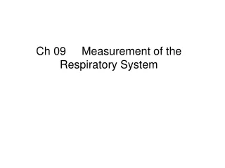
Spirometer Rotational displacement sensor Other signal processing Counterweight Strip-chart recorder Kymograph Bell PS VS TS Water seal Blood flow Uabs Mouthpiece FS x One-way valves Soda-lime canister VL Ubs TL Thermometer for spirometer gas temperature PA FA x QAWO Spirometer system Pulmonary system Figure 9.6 A water-sealed spirometer set up to measure slow lung-volume changes. The soda-lime and one-way-valve arrangement prevent buildup of CO2 during rebreathing.
Soda lime Soda lime is a mixture of chemicals, used in granular form in closed breathing environments, such as general anaesthesia, submarines, rebreathers and recompression chambers, to remove carbon dioxide from breathing gases to prevent CO2 retention and carbon dioxide poisoning. It is made by slaking quicklime (生石灰 ) with concentrated caustic soda (NaOH) solution. The main components of soda lime are : Calcium hydroxide - Ca(OH)2 (about 75%) Water - H2O (about 20%) Sodium hydroxide - NaOH (about 3%) Potassium hydroxide - KOH (about 1%) From Wikipedia, the free encyclopedia
Absolute volume of the lung – how to measure? How to measure? Difficulty in measuring absolute lung volume * complex geometry, irregular * inaccessibility
Absolute volume of the lung – three methods • Methods: • Nitrogen-washout • -- based on static mass balances and washout of a test gas • 2. Helium dilution • -- based on static mass balances and dilution of a test gas • 3. Total body plethysmography – based on dynamic mass balances and gas compression
Absolute volume of the lung Dalton's lawof partial pressures: the total pressure exerted by a gaseous mixture is equal to the sum of the partial pressures of each individual component in a gas mixture
Absolute volume of the lung Nx mole of X only Nx Vx P Nx mole of X and other gases N moles totally N V P
Absolute volume of the lung – Nitrogen-washout estimation 100% O2 One-way valves TS TL VL FSN2 Spirometer FAN2 VS O2 + N2 Nitrogen analyzer + CO2 Figure 9.7 Diagram of an N2 washout experiment The expired gas can be collected in a spirometer, as shown here, or in a rubberized-canvas or plastic Douglas bag. N2 content is then determined off-line. An alternative is to measure expiratory flow and nitrogen concentration continuously to determine the volume flow of expired nitrogen, which can be integrated to yield an estimate of the volume of nitrogen expired.
100% O2 One-way valves TS TL VL FSN2 Spirometer FAN2 VS O2 + N2 Nitrogen analyzer + CO2
Absolute volume of the lung – Helium-dilution estimation Tracer gases: * Properties: nontoxic, insoluble * Examples: He, Ar, Ne
Spirometer Rotational displacement sensor Other signal processing Counterweight Strip-chart recorder Kymograph Bell PS VS TS Water seal Blood flow Uabs Mouthpiece FS x One-way valves Soda-lime canister VL Ubs TL Thermometer for spirometer gas temperature PA FA x QAWO Spirometer system Pulmonary system Figure 9.6 A water-sealed spirometer set up to measure slow lung-volume changes. The soda-lime and one-way-valve arrangement prevent buildup of CO2 during rebreathing.