Download
1 / 40
400 likes | 511 Views
Explore the intricate details of the integumentary system focusing on skin and its derivatives, such as hair, nails, sweat glands, and more. Learn about the regional differences, adnexal structures, and functional aspects of this vital system.

E N D
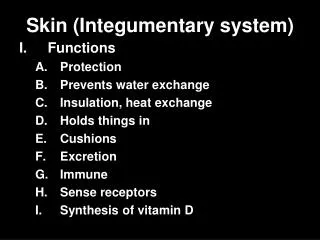
Skin and adnexa • Skin • Adnexa (derivatives): – hair – nail – sebaceous glands – sweat glands – mammary glands
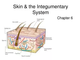
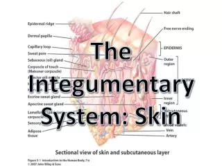
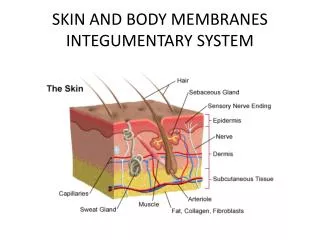
Skin (cutis): structure • epidermis – stratified squamous keratinized epithelium • dermis (corium) –connective tissue + adnexa • hypodermis (tela subcutanea) – dense c.t. + adipocytes (panniculus adiposus) • area 1.5 – 2.5 m2 • weight 16 % of b.w. • Thickness 1 – 5 mm
Stratum corneum propriumthick layer, keratin, desmosome disjunction (Stratum lucidum)cells – no nucleus; tonofilaments, eleidin Stratum granulosumpicnoticcell nuclei , granules with glykolipids and keratohyalin Stratum spinosum„prickle-cell“ or spiny layer, tonofilaments Stratum basalecuboida-columnar cells, mitosis + melanocytes Epidermis(stratif. squamous keratinized ep.) basement membrane
Epidermis Dermis
Epidermis (cells = keratinocytes) stratum corneum stratum granulosum stratum spinosum stratum basale
Another cells in epidermis • A = melanocytes • B = Langerhans‘ cells (antigen presenting) • C = Merkel‘s cells (receptors) „supranuklear cap“ - granules of pigment melanin
Thin 0.5-5.0 mmepidermis70 – 100 m 4 layers, + all adnexa Occurrence: all over body, except in glans penis and labia minora + vestibulum vaginae Thick 0.8-1.5 mm 400 – 800 m,5 layers of epidermis, without hair follicles . Occurrence: - palma manus - planta pedis Types of skin
Skin type: thin thick REGIONAL DIFFERENCES Skin with hair Skin from axilla Skin from tip of finger apocrine sweat glands ONLY ecrine sweat glands NO! YES! hair follicles, sebaceous gll. NO!
Epidermis of thick skin Cristae et sulci cutis: papilary lines (dermatoglyphic patterns = fingeprints) Sweat glands vessels of subpapilar plexus nerv fibers crista intermedia = epidermal peg dermal papillae
Stratum papillaredermal papillae – loose connective tissue + capillary loops and nerve endings Stratum reticulareirregular dense c.t.(dermatansulphate) with solitary smooth muscle cells, + SKIN ADNEXA Dermis
Skin derivatives (adnexa) • Keratinized type: - hair - nail • Non-keratinized type: - sweat gland - sebaceous gland - mammary gland
Hair (pilus) • Scapus • Radix • Bulbus • M. arrector pili • Gl. sebacecea
Hair and hair follicle: Scapus pili Radix pili: kutikula kůra dřeň Zevní kořenová pochva Vnitřní kořenová pochva Vazivová pochva Vazivová papila (papilla pili) epidermis dermis gl. sebacea m. arrector pili Bulbus pili
Hair / pilus dermal sheath (3) outer epithel. sheath (2) inner epithel. sheath (1): Henle Huxley sheath cuticule hair cuticule cortex (dřeň) papilla blood vessels hair (medulla) melanocytes
Skin with hair (HES) m. arrector pili
Skin with hair (HEŠ) Dermal sheath Hair follicle Hair: cortex Bulbus pili Melanocytes Papilla pili
hair spruce-cone Hair cuticle
Nail (unguis) Nail plate (str. corneum) Radix unguis Epithelial bed (str. germinativum) Lectulusunguis kost Periost Kůže bříška prstu
Nail (HE, cross section) Nail plate Epithelial bed Vallum unguis Sulcus unguis Dermal bed (lectulus unguis) Bone of distal phalanga
Skin glands small – tubular eccrine • sweat gland large – tuboalveolarapocrine • sebaceous gland – alveolar holocrine • mammary gland – tuboalveolar
Sweat glandsgll. sudoriferae • Tubular glands: • secretory portion • duct • epidermal canal • Eccrine (eccrinae) – small, • Apocrine (apocrinae) – large, aromatic eccrine apocrine
Sweat glands • duct: in epidermis – intraepidermal canal in dermis – 2layered squamous epithelium • Secretory portion: „glomerulus“ - dark cells - mucopolyssacharides - light cells – H2O and ions - myoepithelial cells - glandular cells - myoepithelial cells Secretion: H20, proteins, Nacl, NH3, urea, uric acid Apocrine glands - occurrence: axilla, cirkumanal region, labia pudendi minores, areola mammae, glands of Moll In palpebra, gll. ceruminosae Secretion: H20, proteins, steroids
Epidermal canal of sweat gland:
duct: stratified squamous ep. Secretory portion – filled with cells Secretion: triacylglycerol, cholesterol, squalene Sebaceous glands alveolar, holocrine
Gl. sebaceae (HE) Pyknotic nucleus Center of alveol Basal cells – proliferating („germinal“) cells
Mammary glandgl. mammae Tuboalveolar gland
Intergumentary system Slides: • 69. Skin from tip of finger (HE) • 70. Skin from axilla (HE) • 71. Skin with hair (HE) • 72. Nail (HE, longitudinal or cross section) • 73. Mamma non lactans (HE) • 74. Mamma lactans (HE)