Download
1 / 43
540 likes | 1.78k Views
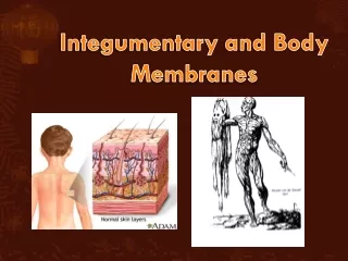
SKIN AND BODY MEMBRANES INTEGUMENTARY SYSTEM. EPITHELIAL MEMBRANES. There are 3 types of epithelial membranes Cutaneous membrane Mucus membrane Serous membrane. Cutaneous Membrane. Skin Dry Membrane. Mucous Membrane. Lines all body cavities that are open to the exterior.

E N D
EPITHELIAL MEMBRANES • There are 3 types of epithelial membranes • Cutaneous membrane • Mucus membrane • Serous membrane
Cutaneous Membrane Skin Dry Membrane
Mucous Membrane Lines all body cavities that are open to the exterior. ex: Respiratory, digestive, urinary, and reproductive Moist membrane
Serous Membrane Lines cavities that are closed to the exterior. Occur in pairs. Reduces friction, especially in organs that move Ex: heart, stomach, lungs (Think of a pushed in balloon. The space between is not filled with air but serous fluid.)
INTEGUMENTARY SYSTEM (SKIN, HAIR, NAILS) • Basic Functions • Synthesizes vitamin D • Excretion of urea and salts • Protects deeper tissue from: • Mechanical damage • Chemical damage • Bacteria • Ultraviolet radiation • Thermal damage • Drying out
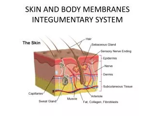
STRUCTURE OF THE SKIN • There are 3 major layers of the skin • Epidermis • Dermis • Hypodermis (subcutaneous tissue/layer)
EPIDERMIS • Outer layer • Made up of squamous epithelium that can be keratinized (hard or tough) • Melanocytes found in deepest layer – gives skin its color. • Mostly dead cells • Avascular – no blood vessels. • Mostly keratinocyte cells. They produce keratin, which is a protein that makes the epidermis a tough layer. • New cells are produced constantly and pushed upward. • New epidermal layer every 2-4 weeks
DERMIS • Found under the epidermis (2nd layer) • Made up of dense connective tissue. • Varies in thickness • The epidermis and dermis are connected but rubbing may cause them to separate resulting in a blister • Structures found in the dermis include: papillary layer, sebaceous glands, sweat glands, hair, hair follicle, erector pilli, blood vessels.
STRUCTURES IN THE DERMIS PAPILLARY LAYER • Upper layer of the dermis. • Has finger like projections called dermal papillae which on the hands and feet are arranged in definite patters that form ridges. • They enhance the gripping ability of the fingers and feet • Patterns are genetically determined & form your fingerprints.
STRUCTURES IN THE DERMIS Sebaceous glands • Also known as oil glands • Keep skin moist and prevents hair from becoming brittle by producing sebum • Contains chemicals that kill bacteria. • When sebum blocks the glands duct, a whitehead appears. • Acne is an infection of the sebaceous glands.
STRUCTURES IN THE DERMIS Sweat Glands • Produce sweat which is acidic (pH 4-6) • Inhibits bacteria and gets rid of excess heat
STRUCTURES IN THE DERMIS Hair & the hair follicle • Hair is produced by a hair follicle. • The root is enclosed by the follicle and the shaft is projecting from the surface. • Cuticle – outer most layer of hair in which cells overlap like layers on a roof. • When the cuticle wears away, “split ends” occur because the inner fibers frizz out.
HYPODERMIS/SUBCUTANEOUS TISSUE • Adipose tissue (Fat) • Shock absorber • Insulates
MELANIN • A pigment that ranges in color from yellow to brown to black. • Produced by cells called melanocytes. • Sunlight causes melanin to be produced & deepest cells of the epidermis. • Melanin forms a protective umbrella over the nucleus of the cells. Protects against ultraviolet radiation from sunlight.
Melanin • Freckles and moles are seen when melanin is concentrated in one spot.
SUN EXPOSURE • Despite melanin’s protection, excessive sun exposure eventually damages skin. • It causes elastic fibers to clump, leading to leathery skin. • Sun exposure also depresses the immune system, can activate cold sores, and alter DNA and lead to skin cancer.
DISCOLORATION OF SKIN CYANOSIS JAUNDICE A yellow coloration to the skin usually signifies a liver disorder in which excess bile pigments are absorbed into the blood. • A bluish coloring of the skin when the blood is poorly oxygenated.
DISCOLORATION OF SKIN BRUISES • Site where blood has escaped from the blood vessels and has clotted in tissue spaces
BURNS A burn is damage to your body's tissues caused by heat, chemicals, electricity, sunlight or radiation. Scalds from hot liquids and steam, building fires and flammable liquids and gases are the most common causes of burns.
1ST DEGREE BURNS First-degree burns, the mildest of the three, are limited to the top layer of skin: Signs and symptoms: These burns produce redness, pain, and minor swelling. The skin is dry without blisters. Healing time: Healing time is about 3 to 6 days; the superficial skin layer over the burn may peel off in 1 or 2 days. Only the epidermis is damaged in 1st degree burns.
2ND DEGREE BURNS Second-degree burns are more serious and involve the epidermis and the upper region of the dermis. Signs and symptoms: These burns produce blisters, severe pain, and redness. The blisters sometimes break open and the area is wet looking with a bright pink to cherry red color. Healing time: Healing time varies depending on the severity of the burn. It can take up to 3 weeks or more. Usually little to no scarring if care is taken to prevent infection.
3RD DEGREE BURNS Third-degree burns are the most serious type of burn and involve all the layers of the skin and underlying tissue: Signs and symptoms: The surface appears dry and can look waxy white, leathery, brown, or charred. There may be little or no pain or the area may feel numb at first because of nerve damage. Healing time: Healing time depends on the severity of the burn. Deep second- and third-degree burns (called full-thickness burns) will likely need to be treated with skin grafts, in which healthy skin is taken from another part of the body and surgically placed over the burn wound to help the area heal.
SKIN CANCER • Basal Cell Carcinoma • Squamous Cell Carcinoma • Malignant Melanoma
SKIN CANCER Basal cell carcinoma • Most common • Least malignant • Cells of the epidermis are altered and no longer form keratin but invade the dermis and subcutaneous tissue. • Slow growing and 99% of the cases are fully cured if surgically removed.
SKIN CANCER Squamous cell carcinoma • Rapid growing • Will spread to lymph nodes if not removed • Epidermal cancer • If removed early, good chance for complete recovery
SKIN CANCER Malignant melanoma • Cancer of melanocytes • Least common • Often deadly • Appears as a spreading (metastasizing) brown to black patch. • Can invade surrounding lymph and blood vessels. • 50% survival rate; early detection increases survival.
Psoriasis Psoriasis is a common skin condition that causes skin redness and irritation. Most people with psoriasis have thick, red skin with flaky, silver-white patches called scales. Psoriasis seems to be passed down through families. Doctors think it probably occurs when the body's immune system mistakes healthy cells for dangerous substances. It is not contagious. Anyone can get it, but it most commonly begins between ages 15 and 35. The following may trigger an attack of psoriasis or make the condition more difficult to treat: Bacteria or viral infections, including strep throat and upper respiratory infections Dry air or dry skin Injury to the skin, including cuts, burns, and insect bites Some medicines, including anti-malaria drugs, beta-blockers, and lithium Stress Too little sunlight Too much sunlight (sunburn) Too much alcohol
Psoriasis continued Symptoms Treatment Skin lotions, ointments, creams, and shampoos. These are called topical treatments. Pills or injections that affect the body's immune response, not just the skin. There are called systemic, or body-wide, treatments. Phototherapy, which uses light to treat psoriasis. • Irritated, red, flaky patches of skin • Most often seen on the elbows, knees, and middle of the body • Red patches may appear anywhere on the body, including the scalp • The skin may be: • Itchy • Dry and covered with silver, flaky skin (scales) • Pink-red in color (like the color of salmon) • Raised and thick • Other symptoms may include: • Genital lesions in males • Joint pain or aching • Nail changes, including thick nails, yellow-brown nails, dents in the nail, and nail lifts off from the skin underneath • Severe dandruff on the scalp
Boils A boil is a skin infection involving an entire hair follicle and nearby skin tissue. They are generally caused by the bacteria Staphylococcus aureus, but they may be caused by other bacteria or fungi found on the skin's surface. Damage to the hair follicle allows these bacteria to enter deeper into the tissues of the follicle and the tissue underneath. A boil may begin as a tender, pinkish-red, swollen, firm area in the skin. Over time, it will feel like a water-filled balloon or cyst.
Boils continued Symptoms Treatment Boils usually must open and drain before they will heal. This usually occurs in less than 2 weeks. Warm, moist compresses help boils drain, which speeds healing. Gently soak the area with a warm, moist cloth several times each day. Never squeeze a boil or try to cut it open at home. This can spread the infection and make it worse. When the boil finally does burst and drain, continue to put warm, wet compresses on the area. Deep or large boils may need to be drained with surgery by a health care provider. • Is usually pea-sized, but may be as large as a golf ball • May develop white or yellow centers (pusteles) • May join with another boil or spread to other skin areas • May grow quickly • May weep, ooze, or crust • Other symptoms may include: • Fatigue • Fever • General ill-feeling • Itching before the boil develops • Skin redness around the boil
Warts Warts are small, usually painless growths on the skin caused by a virus called human papillomavirus (HPV). Most, but not all, are generally harmless. All warts can spread from one part of your own body to another. They may spread from one person to another, but this is uncommon. Different types of warts include: Common warts usually appear on the hands, but can appear anywhere. They usually do not cause pain unless they are repeated rubbed against. Flat warts are generally found on the face and forehead. They are common in children, less common in teens, and rare in adults. Genital warts(condyloma) are usually found on the genitals, in the pubic area, and in the area between the thighs, but they can also appear inside the vagina and anal canal. Plantar warts are found on the soles of the feet. They can be very painful. Many of them on the foot may cause difficulty walking or running. Subungual and periungual warts appear under and around the fingernails or toenails.
Warts continued Symptoms Treatment Do NOT attempt to remove a wart yourself by burning, cutting, tearing, picking, or any other method. Over-the-counter medications are available to remove warts. Do NOT use over-the-counter wart medications on your face or genitals. Warts on the face or genitals need to be treated by your doctor or nurse. Your health care provider may recommend the following treatments if your warts do not go away: Stronger (prescription) medications, such as podophyllin or salicylic acid A blistering solution Freezing the wart (cryotherapy) to remove it Burning the wart (electrocautery) to remove it Laser treatment for difficult to remove warts Immunotherapy, which gives you a shot of a substance that causes an allergic reaction and helps the wart go away Skin medicine called imiquimod • The typical wart is a raised round or oval growth on the skin with a rough surface. • The spot may be lighter, darker, or black (rare) colored compared to other skin. • Some warts have smooth or flat surfaces. • Some warts cause pain, others do not.
Rosacea Rosacea is a chronic skin condition that makes your face turn red and may cause swelling and skin sores that look like acne. Rosacea is a harmless condition, but it may cause you to be self-conscious or embarrassed. The cause is not known. You may be more likely to have this if you are Age 30-50 Fair-skinned A woman (but men usually have more severe symptoms) Rosacea involves swelling of the blood vessels just under the skin. It may be associated with other skin disorders (acne vulgaris, seborrhea) or eye disorders (blepharitis, keratitis).
Rosacea continued Symptoms Treatment There is no known cure for rosacea. Your doctor will help you identify the things that make your symptoms worse. These are called triggers. Avoiding your triggers may help you prevent or reduce flare-ups. Here are some steps that may help ease or prevent symptoms: Avoid sun exposure. Use sunscreen every day. Avoid a lot of activity in hot weather. Try to reduce stress. Try deep breathing, yoga, or other relaxation techniques. Limit spicy foods, alcohol, and hot beverages. Triggers vary from person to person. Other triggers may include wind, hot baths, cold weather, specific skin products, exercise, or other factors. • Redness of the face • Blushing or flushing easily • A lot of spider-like blood vessels (telangiectasia) of the face • Red nose (called a bulbous nose) • Acne-like skin sores that may ooze or crust • Burning or stinging feeling in the face • Irritated, bloodshot, watery eyes
Rosacea continued • Treatment (continued) • Antibiotics taken by mouth (such as tetracycline, minocycline, or doxycycline) or applied to the skin (such as metronidazole) may control acne-like skin problems. • Other medications (isoretinol or Accutane), which are similar to vitamin A, are stronger alternatives that your doctor or dermatologist might consider. • Rosacea is not acne and will not improve with over-the-counter acne treatment. • In severe cases, laser surgery may help reduce the redness. Surgery to remove some swollen nose tissue may also improve your appearance.
Impetigio Impetigo, a skin infection, is caused by streptococcus (strep) or staphylococcus (staph) bacteria. Methicillin-resistant staph aureus (MRSA) is becoming a common cause. It is most common in children, particularly those in unhealthy living conditions. Involves the top layers of the skin. Impetigo is contagious, meaning it can spread to others. You can catch this infection if the fluid that oozes from the blisters touches an open area on your skin. Diagnosis is based mainly on the appearance of the skin lesion. A culture of the skin or lesion usually grows the bacteria streptococcus or staphylococcus. The culture can help determine if MRSA is the cause, because specific antibiotics are used to treat this infection.
Impetigo continued Symptoms Treatment The goal is to cure the infection and relieve the symptoms. A mild infection may be treated with a prescription antibacterial cream. More severe cases may require antibiotics, taken by mouth. Wash (do not scrub) the skin several times a day, preferably with an antibacterial soap, to remove crusts and drainage. • A single or possibly many blisters filled with pus; easy to pop and -- when broken -- leave a reddish raw-looking base (in infants) • Itching blister: • Filled with yellow or honey-colored fluid • Oozing and crusting over • Rash -- may begin as a single spot, but if person scratches, it may spread to other areas • Skin lesions on the face, lips, arms, or legs, that spread to other areas • Swollen lymph nodes near the infection (lymphadenopathy)
Jaundice continued Symptoms Treatment Treatment depends on the cause of the jaundice. • Yellow skin and the white part of the eyes (sclera) -- when jaundice is more severe, these areas may look brown • Yellow color inside the mouth • Dark or brown-colored urine • Pale or clay-colored stools
Jaundice Jaundice is a yellow color of the skin, mucus membranes, or eyes. The yellow coloring comes from bilirubin, a byproduct of old red blood cells. Jaundice can be a symptom of other health problems. Everyday, a small number of red blood cells in your body die, and are replaced by new ones. The liver removes the old blood cells, forming bilirubin. The liver helps break down bilirubin so that it can be removed by the body in the stool. When too much bilirubin builds up in the body, jaundice may result. Jaundice can occur if: Too many red blood cells are dying or breaking down and going to the liver The liver is overloaded or damaged The bilirubin from the liver is unable to move through the digestive tract properly Jaundice is often a sign of a problem with the liver, gallbladder, or pancreas. Infections, use of certain drugs, cancer, blood disorders, gallstones, birth defects and a number of other medical conditions can lead to jaundice.