Download
1 / 37
412 likes | 1.39k Views
Integumentary System: Skin and adnexa. Lecture 4 Tuesday, January 23, 2007 Refs. Ross and Pawlina Histology Chapter 15, Wheater’s Functional Histology Chapter 9 diFiore Atlas of Human Histology Medical Physiology p. 572, 352, 1231-7. Skin. Functions Protection

E N D
Integumentary System:Skin and adnexa Lecture 4 Tuesday, January 23, 2007 Refs. Ross and Pawlina Histology Chapter 15, Wheater’s Functional Histology Chapter 9 diFiore Atlas of Human Histology Medical Physiology p. 572, 352, 1231-7
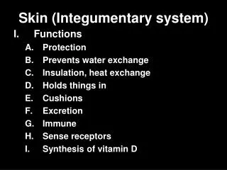
Skin • Functions • Protection • Mechanical, dehydration, UV radiation • Immune system (innate and APC cells) • Largest sensory organ • Thermoregulation • Metabolism • Vitamin D synthesis • Adipose tissue
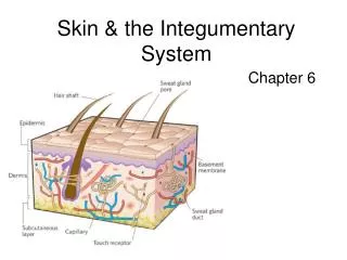
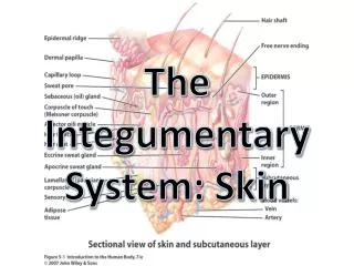
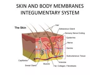
3 Layers of the skin • Epidermis • Keratinizing stratified squamous epithelium • Self renewing, avascular • Dermis • Supporting and nourishing layer • Collagen, elastic fibers, vessels and nerves • Hypodermis or subcutis • Loose CT, adipose cells, variable thickness
Thin skin, full thickness diFiore Plate 46Trichrome stain, low magnification
Epidermal-dermal junction • Epidermal ridges (rete ridges) interdigitate with dermal papillae • Superficial dermis = papillary dermis • Fine collagen and elastic fibers • Vascular plexus • Deep dermis = reticular dermis • Thick collagen and elastic fibers • Larger vessels
Regional variations • Thick skin • Palmar and plantar surfaces (glabrous=hairless) • Epidermis is relatively thicker, keratinized layer is very thick, rete ridges more prominent • Thin skin • Most of body--especially eyelids and periorbital skin • Hair in thin skin is fine. • Scalp skin • Restricted to head • More numerous and larger hair follicles
Upper panel: thin skin (x 123) WFH 9.3 and lower panel thick skin (x104) WFH 9.2 H&E .
Epidermis • 4 layers from dermis to surface • Stratum basale • Germinal layer • Stratum spinosum • Stratum granulosum- keratohyalin granules • Stratum corneum • Keratinocytes= name for epithelial cells in skin • Do not confuse with keratocyte in the cornea. • Basal to desquamation - 25-50 days
Stratum spinosum EM, 25,000x WFH 9.6bD=desmosomeK=keratinocyte
Stratum granulosum EM, 5000x WFH 9.6cK= keratohyaline granules
Dermal-epidermal junction, EM 24000x WFH 9.5bHD= hemidesmosome3 zones of basement membraneL=lamina lucidaD=lamina densaF= fibroreticular lamina
Other cells in the epidermis • Melanocytes • Synthesize and release melanin • Premelanosome-melanosome • Langerhans cells • APC, Birbeck granules • Merkel cells • Touch receptors in basal layer
Langerhans cells and EM of Birbeck granules in cytoplasm of Langerhans cell.WFH 9.7c,d
Adnexa • Also referred to as skin appendages • Derived embryologically from epidermis • Hair follicles • Sweat glands • Merocrine-open onto surface • Apocrine-open into hair follicles • Sebaceous glands-open into hair follicles
Hair follicles • Longitudinal segments • Infundibulum--segment from the skin surface to the level of sebaceous ducts. • Isthmus--segment from sebaceous ducts to insertion of arrector pili muscle. • inferior segment-- ends in expanded hair bulb • Dermal papilla--tuft of connective tissue that invaginates the base of the bulb. • Matrix cells-- cells that surround the dermal papilla and proliferate generating hair.
Concentric layers of the hair and follicle • Medulla (often not present in fine hair) • Cortex- thick , highly keratinized layer • Cuticle-thin, hard, keratinized layer • Internal root sheath • External root sheath • Glassy membrane--thick basement membrane
Hair: types and growth • Hair thickness varies. • Vellus = fine hair • Terminal is coarse as in scalp hair. • Phases of hair growth: • Growing phase is anagen • Period in which growth stops is catagen • Resting phase is telogen • Follicles are shorter. • Hair bulb is smaller and lacks dermal papilla.
Pilosebaceous unit WFH 9.14aM= arrector pili musclesympathetic innervation
Merocrine sweat gland WFH 9.15aSympathetic innervation:Cholinergic fibers
Apocrine sweat gland WFH 9.16Misnomer- secretion is not apocrine.Sympathetic innervation byadrenergic fibers
Thermoregulation • Body core temperature remains almost constant. • Increase in body core temperature causes: • Vasodilation of dermal vessels and increased heat loss from skin. • Evaporation of sweat from surface increases heat loss. • Two types of skin in terms of thermoregulation: apical and nonapical: • Apical skin (nose, ears, fingers and toes) has many glomus bodies; has significant sympathetic tone • Nonapical skin has few, if any, glomus bodies; vessels respond mainly to local mediators.
Skin is the largest sensory organ • Mechanoreceptors • Meissner’s corpuscles in the epidermal ridges • Ruffini’s corpuscles • Pacini’s corpuscles are deep (subcutis) • Merkel’s cells/disks at junction of epidermis and dermis • Krause end bulbs • Chemoreceptors • Nociceptors respond to painful stimuli • Temperature receptors in skin • Respond to local heat by vasodilation or cold by vasoconstriction. • Send message about environment to hypothalamus-anticipatory feedback.