Download

1 / 36
370 likes | 558 Views
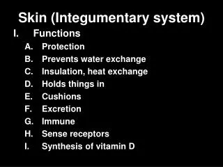
Skin Integumentary system 2009. Skin (cutis, derma ). The heaviest organ in the body - 16% of weight Size: 1,2-2,3m 2 Function: Protection (mechanic, from desiccation) Thermoregulation Sweat glands (perspiratio insensibilis) Changes in blood flow Excretion of salt (iron looses)

E N D
Skin (cutis, derma) • The heaviest organ in the body - 16% of weight • Size: 1,2-2,3m2 • Function: Protection (mechanic, from desiccation) • Thermoregulation • Sweat glands (perspiratio insensibilis) • Changes in blood flow Excretion of salt (iron looses) Nonspecific immunity Metabolism- ergosterol-vit.D Sensoric ending Sexual signaling (Endocrine gland – adipose tissue: leptin, adiponectin, estrogenes)
Development • Ectoderm • Periderm – 2 layers cuboidal and flat superficial • Stratified squamous • Glands, hair and nails – invagination of ectoderm into dermis
Skin lines (lineae distractiones) • Externally visible skin lines: • Wrinkle lines: lines of expression • Flexure (joint) lines • sulcus mentolabialis, nasolabialis • sulcus gluteus
Surface pattern lines - hand – Purkyně - chiromantia • linea oppositionis pollicis (vitalis) • linea manus clausae (cephalica, naturalis) • linea occlusionis dig. trium ulnarium (mensalis) • sulcus cutaneus intercarpalis (linea rasceta) • linea restricta
Skin • Grooves(sulci cutis) • Papillary ridges (friction ridges)(cristae cutis) – dermatoglyphics - 9 types (according to Purkyně) → daktyloscopy – forensic importance • Toruli tactiles– 10 in hand ( thenar) • Lineae distractiones • Retinacula cutis - (retinaculum caudale) • Intristinc scarring - Striae cutaneae – rupture of lateral cohesion of the collagen fibres -growth, pregnancy and obesity
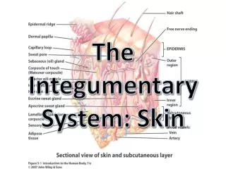
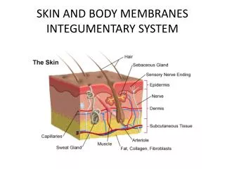
Skin • Epidermis • Dermis • Hypodermis (tela subcutanea – panniculus adiposus) • Appendages: • Hairs • Nails • Glands of the skin • Mammary gland
Epidermis • Stratified squamous keratinized epithelium + melanocytes, Langerhans cells, Merkel´s cells • Thick type: palms of hands and soles of the feets • Thin type: rest of the body Cell renewal in epidermis:15 – 30 days EGF, Keratinocyte growth factor, retinoic acid (vit. A)
Epidermis • Stratum basale (basophilic cuboid to columnar cells on lamina basale – stem cells) • Stratum spinosum – polygonal cells, contain of cytokeratin filament, desmosomes • Stratum granulosum – 3 – 5 layers of cells – keratohyalin granules – basophilic; lamellar granules – lipids (ceramids) – barrier – sheet containing lipid • Stratum lucidum – in thick type -eosinophilic • Stratum corneum – cells filled by keratin filament are packed together by filaggrin; desmosomes
Stem cells • Stem cells in the bulb region of the hair follicle – indepedent migration: • Bulb-hair stem pathway – at the apex of the dermal papilla • Bulb-epidermis cell pathway – epidermis – stratum basale • Sebaceous glands
Differentiation of a keratinocyte • Cells of stratum spinosum – synthesis of acylglucosylceramide – RER + GA = membrane-coating granules (or lamellar bodies) • Bb. stratum granulosum – proteosynthesis = keratohyaline granules, lamellar bodies release ceramide into the intercellular spaces • Bb. stratum lucidum– intermediate layer - eosinophilic • Bb. stratum corneum – without nuclei, keratin croslinked by filaggrin, together with ceramide form cell envelope, cells are joined by desmosomes
Melanocytes • Development from neural crest • Eumelanin, pheomelanin (contains cysteine) - red hair • Tyrozine – dopa – dopaquinone – melanin (tyrozinase) • Synthesis:vesicle with enzymatic activity, fine granular material • Melanosome – filaments with periodocity 10nm • Dense granule • Melanosome visible in LM, size 1x0,4 m
Melanocytes • Cytocrine secretion melanosomes transferred to keratinocytes • Function – protection from UV radiation • Epidermal – melanin unit – about 1000/mm2 • Higher number in the skin of scrotum, circum-anal region, areola mammae)
Merkel´s cells • Present in thick skin in stratum basale • Small dense granules (neurotransmiters) • Nerve ending • Sensoric mechanoreceptors
Langerhans´ cells • Mainly in stratum spinosum • Bone marrow derived - antigen presenting cells – in lymphatic nodes - they differentiate into activated dendritic cells – (contain Birbeck´s or vermiforms granules - rodlike)
Dermis • Dense collagen connective tissue with elastic fibres. Main glycosaminoglycane is dermatan sulphate • Attachment to epidermis – hemidesmosomes and anchoring filaments (laminin 5) and fibriles (collagen VI) – blister, pemphigus • Stratum papillare • Stratum reticulare • Skin appendages – glands, hairs, nails • Sensoric ending (Vater-Paccini, Meissner etc.)
Fissionability lines • Along the collagen fibres in dermis • Important for cosmetic surgery and for cutting motion podle Kraisla
HypodermisTela subcutanea • Loose collagen tissue and adipose tissue • Is not present in eyelids, clitoris and penis • Retinacula cutis • Panniculus adiposus • Stratum musculorum • Stratum fibrosum • Stratum membranosum • Textus connectivus laxus • Bursae synoviales subcutaneae
Skin appendages • Hair and hair follicle • Sebaceous glands • Sweat gland • Apocrine glands
Hair • Hair follicle • Hair bulb (bulbus pili) • Dermal papilla • Hair (cuticle of hair, cortex, and medulla) • Internal (epithelial) root sheat • External (epithelial) root sheat • Connective tissue sheat • Arrector pili muscle
Nails • Nail root hidden in nail groove • Eponychium or cuticle • Nail plate (stratum corneum) on the nail bed (stratum basale and spinosum) • Nail plate arises from nail matrix (root and lunula)
Glands of the skin • Sebaceous glands– holocrine glands, composed alveolar • Duct – stratified squamous epithelium • Not present in thick type • Duct usually ends in the upper part of hair follicle • Directly on surfacer: glans penis, clitoris, labia minora, lips, areola mammae
Glands of the skin • Sweat glands - eccrine (merocrine) – simple coiled tubular gland • Excretory duct opens at the skin surface • Dark cells – glycoproteins • Clear cells – glycogen, basolateral labyrinth – secretion of water and ionts (Na, Cl) • Myoepithelial cells • Ducts - pseudostratified epithelium and space between keratinocytes • Function - thermoregulation
Glands of the skin • Apocrine glands – present in axilla, anal and genitregion, areola mammae, modified in ear (ceruminous) and eyelid (glands of Moll) • Secretory part is wider, ducts opens in hair follicle, secretory activity starts at puberty (merocrine secretion with changes of cell size) • Adrenergic inervation
Mammary gland (glandula mammaria) • The greatest gland of the skin • Lactation → newborn nutrition • Paired glands – sulcus intermammarius • From 3th – 6th ribs, parasternal ► anterion axillar line • Upon the deep pectoral fascia • Retromammary space – loose connective tissue • 11 cm x 12 cm • 150g, during lactation 300-800g
Development • Mammary ridge Only in thoracal region (Supernumerary breasts and nipples) • Development of nipple and glands • 15-20 lobes epithelial buds – lactiferous ducts • Before puberty lactiferous ducts and lactiferous sinuses, only • Increase in size after puberty (influence of estrogens) - terminal interlobular ducts + adipose tissue
Mammary gland (glandula mammae) • Corpus mammae – glandular tissue - processus axillaris • 15-20 compound tubo-alveolar glands • Ducts = lactiferous ducts → lactiferous sinuses → nipple (mammary papilla) and areola • Lobes → lobules →glandular alveoles surrounded by dense connective tissue and adipose tissue • Fibrous condensations of stromal tissue -to the dermis suspensory ligaments (of Astley Cooper) Retinaculum cutis mammae = ligg. suspensoria mammaria Cooperi
Mamma • Menstrual cycle Progesteron stimulates cyclic changes – alveolar buds develop under the influence of progesteron, old regress (apoptosis) • Pregnancy – Prolactin and placental lactogen – development of secretory acini
Lactation • Nerve stimulus – oxytocin – contraction of myoepithelial cells – rejection of milk • Prolactin – milk secretion • Colostrum -první mléko • Milk – • Proteins – merocrine secretion • Lipids -apocrine secretion • Sugar (lactosa) production in GA • Immunoglobulins (IgA) -plasma cells • Decrease of prolactin level – apoptosis - regression
Mammary gland – arteries • Arcus aortae→ a. subclavia → a. thoracica interna Internal thoracic artery • aa. intercostales anteriores (I.-V./VI.) → rr. perforantes • Arcus aortae→ a. subclavia → a. axillaris Axillary artery • → a. thoracica superior • → (r. pectoralis a. thoracoacromialis, a. thoracica lat., a. subscapularis) • Aorta thoracica • aa. intercostales posterioresIntercostal arteries (II.-V.) → rr. perforantes /II.
Mammary gland – veins • Circular venous plexus Halleri – around areaola • v. axillaris • v. internal thoracic • vv. intercostal
Mammary gland - nerves • nn. intercostales IV.- VI. • rami ant. + lat. • sensoric (T4) and sympathetic fibres
Mammary gland – lymph vessels • 4 quadrants • Plexus subareolaris Sappeyi • Contralateral breast and axilla • Internal mammary lymph node chain • nodi l. parasternales • nodi. mediastinales ant. • nodi epigastrici sup. + inf. • Axillary lymph nodes • Sorgius – The most cranial from nodi l. pectorales,on 2./3. dens of m. serratus ant. • nodi l. supraclaviculares
Carcinoma of mammary gland • The most frequent tumor in females - 9% women suffer from this illness Clinical signs – swelling -tumor, skin pulling, ulcers Examination – palpation, sonography, mamography, lymphatic nodes • exstirpation • mastectomy (parcial, total)
Carcinoma of mammary gland • 90% of carcinoma develop from ductal epithelium, only 10% within lobular alveolo-ductal epithelium • Ductal epithelium has estrogen receptors • Paget carcinoma – in the nipple • Intraductal– within lactiferous ducts • Lobular carcinoma