Download
1 / 1
10 likes | 157 Views
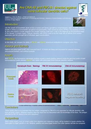
Follicular dendritic cells related to nerve fibres and cellular prion protein expression in ileal and jejunal Peyer’s patches of cows and calves. D. ZIF. GC. GC. SAF84. 6H4. GC. ML. D. AP. SAF34. SAF34. Germinal centre innervation in PP of cows.

E N D
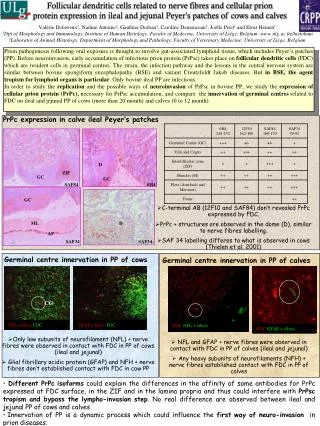
Follicular dendritic cells related to nerve fibres and cellular prion protein expression in ileal and jejunal Peyer’s patches of cows and calves . D ZIF GC GC SAF84 6H4 GC ML D AP SAF34 SAF34 Germinal centre innervation in PP of cows Germinal centre innervation in PP of calves GFAP+ fibres FDC FDCNFL + fibres NFL+ fibres FDC CG FDCGFAP + fibres • Only low subunits of neurofilament (NFL) + nerve fibres were observed in contact with FDC in PP of cows (ileal and jejunal) • Glial fibrillary acidic protein (GFAP) and NFH + nerve fibres don’t established contact with FDC in cow PP • NFL and GFAP + nerve fibres were observed in contact with FDC in PP of calves (ileal and jejunal) • Any heavy subunits of neurofilaments (NFH) + nerve fibres established contact with FDC in PP of calves Valérie Defaweux1, Nadine Antoine2, Gauthier Dorban1, Caroline Demonceau1, Joëlle Piret1 and Ernst Heinen1 1Dpt of Morphology and Immunology, Institute of Human Histology, Faculty of Medecine, University of Liège, Belgium–www.ulg.ac.be/histohum 2Laboratoy of Animal Histology, Department of Morphology and Pathology, Faculty of Veterinary Medecine, University of Liège, Belgium Prion pathogenesis following oral exposure is thought to involve gut-associated lymphoid tissue, which includes Peyer’s patches (PP). Before neuroinvasion, early accumulation of infectious prion protein (PrPsc) takes place on follicular dendritic cells (FDC) which are resident cells in germinal centres. The strain, the infection pathway and the lesions in the central nervous system are similar between bovine spongiform encephalopathy (BSE) and variant Creutzfeldt Jakob diseases. But in BSE, the agent tropism for lymphoid organs is particular. Only bovine ileal PP are infectious. In order to study the replication and the possible ways of neuroinvasion of PrPsc in bovine PP, we study the expression of cellular prion protein (PrPc), necessary for PrPsc accumulation, and compare the innervation of germinal centres related to FDC on ileal and jejunal PP of cows (more than 20 month) and calves (0 to 12 month). PrPc expression in calve ileal Peyer’s patches • C-terminal AB (12F10 and SAF84) don’t revealed PrPc expressed by FDC. • PrPc + structures are observed in the dome (D), similar to nerve fibres labelling. • SAF 34 labelling differes to what is observed in cows (Thielen et al. 2001) • Different PrPc isoforms could explain the differences in the affinity of some antibodies for PrPc expressed at FDC surface, in the ZIF and in the lamina propria and thus could interfere with PrPsc tropism and bypass the lympho-invasion step. No real difference are observed between ileal and jejunal PP of cows and calves • Innervation of PP is a dynamic process which could influence the first way of neuro-invasion in prion diseases.