Download
1 / 28
551 likes | 3.08k Views
Antigen Presentation and Dendritic Cells . Outline: 1. Review of MHC Class I and MHC Class II Presentation 2. Overview of Dendritic Cells Dendritic Cell Paradoxes Cross Presentation Problems in Cross Presentation Pathogens Exploit DCs as an Entry Route for Infection

E N D
Antigen Presentation and Dendritic Cells Outline: 1. Review of MHC Class I and MHC Class II Presentation 2. Overview of Dendritic Cells • Dendritic Cell Paradoxes • Cross Presentation • Problems in Cross Presentation • Pathogens Exploit DCs as an Entry Route for Infection • Pathogens Block Antigen Presentation • Additional Reading J. R. Lingappa, Pabio 552
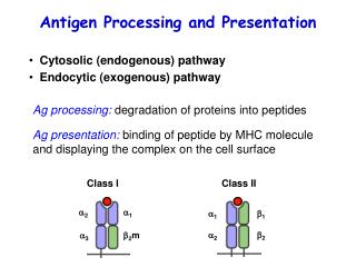
1. Review of MHC Class I and MHC Class II Presentation: Antigen Presentation and Dendritic Cells A. How T cells are primed to recognize microbial antigens: MHC I vs. MHC II presentation Intracellular antigens (typically viral) presented via MHC I on antigen presenting cells (APC) to CD8+ (cytotoxic) T cells. Note: MHC class I expressed on all cell types. Intravesicular antigens (typically bacterial or parasitic) presented via MHC II on APC to CD4+ T cells From Janeway, Immunobiology, 5th edition J. R. Lingappa, Pabio 552
Antigen Presentation and Dendritic Cells Viral antigens from cytosol are presented by MHC class I 1. Review of MHC Class I and MHC Class II Presentation, cont.: A. MHC class I presentation: From Janeway, Immunobiology, 5th edition J. R. Lingappa, Pabio 552
Antigen Presentation and Dendritic Cells MHC class I chains are retained in the ER until peptide binds 1. Review of MHC Class I and MHC Class II Presentation, cont.: A. MHC class I presentation, cont.: From Janeway, Immunobiology, 5th edition J. R. Lingappa, Pabio 552
Antigen Presentation and Dendritic Cells Review of MHC Class I and MHC Class II Presentation, cont.: B. MHC class I presentation results in binding of T cell receptor and activation of CD8+ (cytotoxic) T cells: From Janeway, Immunobiology, 5th edition J. R. Lingappa, Pabio 552
Antigen Presentation and Dendritic Cells Endocytosed bacterial antigens presented by MHC class II 1. Review of MHC Class I and MHC Class II Presentation, cont.: C. MHC class II presentation: From Janeway, Immunobiology, 5th edition J. R. Lingappa, Pabio 552
Antigen Presentation and Dendritic Cells Invariant chain prevents other proteins in the ER from binding to MHC class II Review of MHC Class I and MHC Class II Presentation, cont.: C. MHC class II presentation, cont.: From Janeway, Immunobiology, 5th edition J. R. Lingappa, Pabio 552
Antigen Presentation and Dendritic Cells Review of MHC Class I and MHC Class II Presentation, cont.: D. MHC Class II presentation on APC’s activates CD4+ T cells: From Janeway, Immunobiology, 5th edition J. R. Lingappa, Pabio 552
Antigen Presentation and Dendritic Cells 1. Review of MHC Class I and MHC Class II Presentation, cont.: E. Activation of naïve T cells requires two signals (“co-stimulation”). 1. Signal 1: recognition of peptide-MHC complexes by T cell receptor. 2. Signal 2: interactions between B7(CD80 or CD86) on the APC cell surface and CD28 receptors on the T cell. B7:CD28 binding is promoted and sustained by CD40 binding to CD40 ligand. ONLY PROFESSIONAL APCS CAN CO-STIMULATE AND ACTIVATE NAÏVE T CELLS. PROFESSIONAL APCS ONLY EXPRESS B7 WHEN ACTIVATED BY INFLAMMATION. J. R. Lingappa, Pabio 552
Antigen Presentation and Dendritic Cells 3. Overview of Dendritic Cells: Basic Concepts: 1. DCs are the most effective professional APCs -Only professional APCs are capable activation of naïve T cells -Amateur APCs lack the ability to elicit signal 2 and therefore only recognize previously activated T cells. Most cells that become infected are amateur APCs. -DCs: most effective because of costimulation efficiency, wide distribution, location at critical sentinel sites, and ability to migrate -Activated T cells in turn activate B cells, T cells, and macrophages -Thus, DCs initiate immune responses that are conducted by other cell types. J. R. Lingappa, Pabio 552 From Janeway, Immunobiology, 6th edition
Antigen Presentation and Dendritic Cells 2. Overview of Dendritic Cells, cont.: A. Basic Concepts, cont.: 2. DCs exist in 2 functionally different states (mature and immature). DCs were initially thought to be good phagocytes and weak APCs. Change in our understanding of DC: observation that freshly isolated Langerhans cells were initially weak APCs, but after culture for 1-3 days in presence of antigen they become capable of presentation (due to maturation). Immature and mature DC’s function differently. 3. DCs link the innate and adapative immune response. As immature cells, they act as sensors of the environment. Upon stimulation, dendritic cells undergo maturation and migration to secondary lymphoid organs where they stimulate naïve T cells that then go on to activate T cells, B cells, and macrophages. DC’s are the link between the innate and adaptive immune response. J. R. Lingappa, Pabio 552
2. Overview of Dendritic Cells, cont.: B. Immature Dendritic Cells: Sentinels Antigen Presentation and Dendritic Cells Specialize in endocytosis (antigen uptake) including recycling of MHC I&II Express low levels of MHC class I and MHC class II molecules on cell surface Short half life of MHC class II on surface Rich in vesicles with lysosomal markers that are full of MHC II and HLA-DM called MHC class II compartments or MIIC Express low levels of co-stimulatory molecules on cell surface Antigens taken up are retained for use as immunogenic peptides days later g. Peripheral location: blood, lymphoid, & non-lymphoid tissues (skin &mucosa) From Mellman and Steinman, Cell 106: 255 (2001) J. R. Lingappa, Pabio 552
Antigen Presentation and Dendritic Cells 2. Overview of Dendritic Cells, cont.: C. The Process of Dendritic Cell Maturation: Step 1: DCs sense pathogens or inflammatory signals. i. Microbial products ii. Pro-inflammatory cytokines Maturation occurs only upon detection of microbial products or cytokines. Links efficient antigen presentation to a danger signal: favors activation of T cells ONLY in presence of a pathogen. Step 2: immature DC’s capture the microbe that has been sensed via: i. Phagocytosis (for particulate antigens) ii. Receptor-mediated endocytosis iii. Macropinocytosis (for soluble antigens) Step 3: Migration of DC’s from site of infection towards T cell areas of the proximal LN via afferent lymphatics. Upregulation of the chemokine receptor CCR7 on DC which drives DC migration to: 1. lymphatic vessels by binding 6Ckine released by lymphatic endothelial cells 2. draining lymph nodes (DNL) by binding MIP 3 J. R. Lingappa, Pabio 552
Antigen Presentation and Dendritic Cells 2. Overview of Dendritic Cells, cont.: C. The Process of Dendritic Cell Maturation: Step 4: Decreased endocytic & phagocytic capability Due to down-regulation of macropinocytosis & phagocytosis Mechanism: reduced activation of Cdc42 & Rac (GTPases that regulate actin). Clathrin-mediated endocytosis continues Step 5: Increased surface expression of MHCII bound to peptide Little or no increase in mRNA and protein synthesis Almost entirely due to post-translational events, especially intracellular transport In immature DCs, MHC II accumulates in late endosomes and lysosomes In mature DCs, MHC II accumulates at cell surface Step 6: Enhanced expression of co-stimulatory molecules B7 (CD80 and CD86, CD40) and Fas Step 7: Release of cytokines and chemokines by DCs Including IL-12, IL-18, IL-10 Cytokines polarize T cell responses (see Responses to DCs, below) J. R. Lingappa, Pabio 552
Antigen Presentation and Dendritic Cells 2. Overview of Dendritic Cells, cont.: D. Mature DC’s: Antigen Presenting Cells a. Reduced capacity for antigen uptake b. Increased antigen presentation and T cell stimulation (better than macrophages) c. Redistribution of MHC II from intracellular compartments to cell surface d. MIICs replaced by CIIVs where peptide loading occurs e. CIIVs deliver peptide-loaded MHC II to cell surface f. Dramatic increase in cell surface expression of co-stimulatory molecules g. MHC II loaded with peptide at cell surface has a much longer half life h. Surface expression of co-stimulatory molecules i. Long dendritic processes (membrane folds) for capture of T cells j. Chemokine receptor expression for migration & homing to lymph nodes From Mellman and Steinman, Cell 106: 255 (2001) MIIC J. R. Lingappa, Pabio 552
Antigen Presentation and Dendritic Cells 3. Dendritic Cell Paradoxes: 1. How are 9 - 16 amino acid peptides (ideal for antigen presentation) produced? Lysosomes normally degrade proteins to single amino acids. DC’s regulate lysosomal proteolysis in a developmentally-linked fashion. This may occur in endosome-like vesicles that are specialized for antigen processing. Macrophages contain high levels of proteases resulting in rapid and complete degradation. In contrast, DC’s are protease poor with limited capacity for proteasomal degradation. Dellamare et al., Science 307: 1630, 2005 Limited lysosomal proteolysis favors antigen presentation. J. R. Lingappa, Pabio 552
Antigen Presentation and Dendritic Cells 3. Dendritic Cell Paradoxes: 2. How do DC’s present antigen that was sequestered in lysosomes for days? Lysosomal enzyme content, pH, and morphology are regulated in DC’s. A. Regulation of post-translational trafficking of MHC II: 1. Regulation of cathepsin S Cathepsin S cleaves Ii chain that occupies the MHC II peptide binding groove. Immature DCs: cystatin C antiprotease slows Ii cleavage - MHC II goes to lysosomes. Mature DC’s: cystatin C activity decreases, cathepsin S activity increases and more MHC II reaches the cell surface. 2. Regulation of vacuolar ATPase Low pH favors peptide loading onto MHC II DC maturation lowers endosomal pH Low pH results from recruitment of cytoplasmic ATPase subunits forming functional ATPase Trombetta et al. Science 299: 1400, 2003. (Helicobacter and MTb have ways to block the reduced endosomal pH) J. R. Lingappa, Pabio 552 Trombetta and Mellman, Annual Reviews of Immunology, 23:975, 2005
Antigen Presentation and Dendritic Cells 3. Dendritic Cell Paradoxes: 2. How do DC’s present antigen that was sequestered in lysosomes for days? Lysosomal enzyme content, pH, and morphology are regulated in DC’s. B. Regulation of lysosomal morphology: Time lapse confocal images reveal that within 30 min after stimulation by LPS, lysosomal structures form tubules that take MHC II to plasma membrane. Thus, dendritic cells can rescue components from degradation in lysosomes and transfer them to the plasma membane. Not blocked by MT inhibitors. Formation of tubules may be a specialized function of DC lysosomes. Boes et al., Nature 418: 982, 2002; Chow et al., Nature 418: 988, 2002. Immature (a), maturing (b), and mature (c) dendritic cells stained for MHC class II (green) and lysosomal marker Lamp-1 (red). From Mellman et al., TICB 8: 231 (1998) J. R. Lingappa, Pabio 552
Antigen Presentation and Dendritic Cells 4. Cross-presentation: Do Class I and Class II Presentation explain antigen presentation adequately? J. R. Lingappa, Pabio 552
Antigen Presentation and Dendritic Cells 4. Cross-presentation: Do classical MHC class I and class II Presentation explain antigen presentation fully? Problem 1: Classical MHC class I presentation would require DC’s to get infected and produce peptides in the DC cytoplasm. However, many viruses do NOT infect dendritic cells and still activate cytotoxic CD8+ T cells. There must be a way that dendritic cells can use intracellular peptides produced in other cells to activate cytotoxic T cells. Problem 2: Phagocytosed pathogens such as Salmonella, Brucella, and Leischmania can elicit MHC class I-dependent cytotoxic CD8+ T cell proliferation. To elicit Class I responses, pathogens in phagosomes must transfer antigens into the cytosol. Problem 3: Vaccine antigens are extracellular and yet result in cytotoxic CD8+ T cell responses. Extracellular antigens must be capable of transfer into the cytosol to elicit class I responses. J. R. Lingappa, Pabio 552
Antigen Presentation and Dendritic Cells Sources of antigen for MHC I presentation: Sources of antigen for MHC II presentation: 4. Cross-presentation: Typically MHC II antigens are extracellular and MHC I antigens are cytosolic. However, this is no longer absolutely true - examples that violate both directions exist. Thus, ANY protein can potentially be presented by MHC I or MHC II. Cross presentation = MHC I presentation of exogenous antigens. Trombetta and Mellman, Annual Reviews of Immunology, 23:975, 2005 J. R. Lingappa, Pabio 552
Antigen Presentation and Dendritic Cells 5. Problems in Cross-presentation: Another topological barrier is crossed if extracellular proteins access the proteasome What channel is used when extracellular peptides cross the phagosome membrane? What is the source of the phagosome membrane? J. R. Lingappa, Pabio 552
Exogenous antigens are taken up into a phagosomal compartment composed mainly of PM/endosomes OR Exogenous antigens are moved from phagosome to ER In some organelle (?Phagosome, ? ER-phagosome), retrograde transport to the proteasome in the cytosol occurs. • Peptides produced by this phagosome-associated proteasome are transported back into the phagosome via a TAP transporter and loaded onto MHC class I. Antigen Presentation and Dendritic Cells 5. Problems in Cross-presentation: 2. Models for Cross-Presentation: From Ackerman and Cresswell, Nature Immunology 2004 Jul;5(7):678-84. J. R. Lingappa, Pabio 552
Antigen Presentation and Dendritic Cells A. DC-SIGN captures and transfer HIV-1 DC-SIGN: Adhesion receptor on DCs Binds ICAM-2 promoting trans-endothelial migration of DCs Initiates interaction of DCs with naïve T cells through ICAM-3 binding Binds HIV-1 envelope glycoprotein (gp120) Rather than facilitating infection of DC with HIV, DC-SIGN protects HIV by sequestering it into a protease resistant compartment. DC-SIGN then transfers HIV-1 to CD4+ cells causing infection of these cells. DC-SIGN enhances infection of T cells: at low virus titers T cells not infected unless DCs are present (expressing DC-SIGN in trans). DC-SIGN may transmit other viruses such as Ebola, CMV, and HCV. 6. Pathogens exploit dendritic cells as an entry route for infection: While dendritic cells use pathogen recognition and uptake to activate the adaptive immune system, pathogens conversely use exploit dendritic cells as a route of entry for infection. From Kyook et al. Trends in Mol. Medicine 9:163 (2003) J. R. Lingappa, Pabio 552
Antigen Presentation and Dendritic Cells Fig. 1: Virus captured at surface. HIV-green; HLA - red Fig. 2: Recruitment to cell contact sites. virus-green; mitoch-red. Seen even after protease treatment suggesting internalization Fig. 4: Localization of HIV at antigen-indep. contacts 6. Pathogens exploit dendritic cells as an entry route for infection, cont.: A. DC-SIGN captures and transfer HIV-1, cont. HIV co-opts antigen-capture function of immature DC’s to recruit virus into infectious synapses that contact the T cell McDonald et al., Science 300:1295, 2003 J. R. Lingappa, Pabio 552
1. Escape of viral proteins from proteasomal cleavage. Gly-Ala repeat in EBNA-1 inhibits proteasomal proteolysis. 2. Blockade of TAP-transporter. CMV US-6 inhibits TAP by blocking ATP binding to TAP without affecting peptide binding to TAP. HSV ICP47 blocks peptide-binding site of TAP. 3. Impaired transport of MHC I. CMV US3 binds to 2-microglobulin and causes retention of MHC I-peptide complex in ER. 4. Inhibition of Tapasin. Adenovirus E3-19k binds to MHC class I and TAPand inhibits tapasin causing delayed MHC I maturation. Antigen Presentation and Dendritic Cells 7. Pathogens block antigen presentation: Impairment of MHC class I trafficking by viral gene products: From Yewdell and Hill, Nature Immunol. 3: 1019 (2002) J. R. Lingappa, Pabio 552
Degradation of MHC I. CMV US 2, US 11: membrane glycoproteins that degrade MHC I by promoting ERAD. HIV-1 Vpu induces degradation of CD4 (which needs to bind to MHC class II for T cell activation) via ERAD. 6. Diversion to the lysosome. Murine CMV Gp48 and HHV7 U21 bind to MHC I in ER and divert it to lysosomes for degradation. 7. Downregulation of MHC class I on cell surface. HIV Nef: links MHC I (and CD4) to clathrin (see PM lecture). HHV8 K3 protein induces rapid endocytosis of MHC I. 8. Transcriptional effects (not shown). Adenovirus down-regulates MHC class I transcription via a transcriptional repressor. Antigen Presentation and Dendritic Cells 7. Pathogens block antigen presentation: F. Impairment of MHC class I trafficking by viral gene products, cont.: From Yewdell and Hill, Nature Immunol. 3: 1019 (2002) J. R. Lingappa, Pabio 552
Antigen Presentation and Dendritic Cells 8. Additional Reading (Optional): General Reviews: 1. Mellman, I. And Steinman, R. M. Dendritic cells: specialized and regulated antigen processing machines. Cell 106: 255 (2001) 2. Chow AY, Mellman I. Old lysosomes, new tricks: MHC II dynamics in DCs.Trends Immunol. 26(2):72-8, 2004. 3. Trombetta ES, Mellman I. Cell biology of antigen processing in vitro and in vivo.Annu Rev Immunol. 23:975-1028, 2005. 4. Yewdell J and Hill A. Viral interference with antigen presentation.Nat Immunol. 2002 Nov;3(11):1019-25. Review. Cross Presentation Reviews: 1. Shen L, Rock KL. Priming of T cells by exogenous antigen cross-presente on MHC class I molecules. Curr Opin Immunol. 2006 Feb;18(1):85-91. 2. Touret N, Paroutis P, Grinstein S. The nature of the phagosomal membrane: endoplasmic reticulum versus plasmalemma. J Leukoc Biol. 2005 Jun;77(6):878-85. 3. Gagnon E, Bergeron JJ, Desjardins, M mediated phagocytosis: myth or reality?J Leukoc Biol. 2005 Jun;77(6):843-5. Primary Articles: 1. McDonald, D. et al., Recruitment of HIV and its receptors to dendritic cell-T cell junctions. Science 300:1295 (2003) 2. Chow, A. et al. Dendritic cell maturation triggers retrograde MHC class II transport from lysosomes to the plasma membrane. Nature 418: 988 (2002). 3. Houde M et al. Phagosomes are competent organelles for antigen cross-presentation.Nature. 2003 Sep 25;425(6956):402-6. 4. Gagnon E et al., Endoplasmic reticulum-mediated phagocytosis is a mechanism of entry into macrophages.Cell. 2002 Jul 12;110(1):119-31. 5. Trombetta, E et al. Activation of lysosomal function during dendritic cell maturation.Science. 2003 Feb 28;299(5611):1400-3. 6. Delamarre L, Pack M, Chang H, Mellman I, Trombetta ES. Differential lysosomal proteolysis in antigen-presenting cells determines antigen fate. Science. 2005 Mar 11;307(5715):1630-4. 7. Touret et al., Quantitative and dynamic assessment of the contribution of the ER to phagosome formation.Cell. 2005 Oct 7;123(1):157-70. J. R. Lingappa, Pabio 552