Download
1 / 13
130 likes | 358 Views
Electrocardiography. Prof. K. Sivapalan. Principle of Electrocardiogram. Trunk as volume conductor. Positively charged and negatively charged areas create electrical field. The field changes during spread of impulse. Depolarization towards – upward deflection & away – downward deflection.

E N D
Electrocardiography Prof. K. Sivapalan
Principle of Electrocardiogram. • Trunk as volume conductor. • Positively charged and negatively charged areas create electrical field. • The field changes during spread of impulse. • Depolarization towards – upward deflection & away – downward deflection. ECG
Einthovens triangle. • When the source of the electrical field is at the centre of an equilateral triangle, the sum of electrical potential at the three angles is zero. • Since the heart is roughly at the centre of the triangle and the source moves, 5000 ohm resistance is added to the three leads to get zero potential. ECG
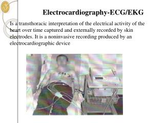
Electrocardiogram. • Electrocardiogram is the record of the changes in the electrical field in the body produced by the electrical activity of the heart. • It represents the sequence and strength of the electrical events in the heart. ECG
Recording ECG. Coronal plane: • Limb leads [Bipolar leads]. • Augmented limb leads.[unipolar] Transverse plane: • Chest [Unipolar] leads V1 to V6 across the chest. Lead I aVR aVL Lead III Lead II aVF ECG
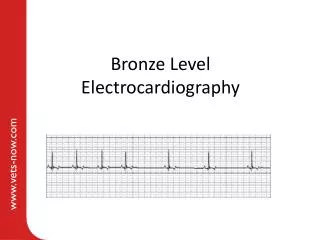
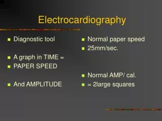
Typical Electrocardiogram. • Paper speed- 25 mm/sec. • Vertically, 1 mV = 10 mm. • P wave – first wave. • Q wave – first down. • R wave – first up in QRS. • S wave – down after R. • T wave – after QRS. • U wave – after T. • PR interval – 0.18 sec. • QRS duration – 0.08 sec. ECG
Information from ECG. • Rate. • Rhythm • Pattern of spread of impulse and integrity of conducting system. • Size [thickness] of myocardium. • Electrolyte imbalance. • Damage to myocardium. ECG
Examples of abnormal ECG. SA node block ECG
Examples of abnormal ECG. Normal. K+ – 4 – 5.5 meq / L K+ – 7 meq / L. K+ – 8.5 meq / L K+ – 3.5 meq /L. K+ – 2.5 meq / L. ECG