Download
1 / 38
410 likes | 942 Views
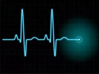
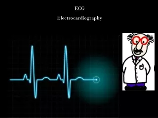
Electrocardiography (ECG, EKG). A typical electrocardiogram: important deflections and intervals. Cardiac Action Potential 과 EKG 와의 관계. P: atrial depolarization QRS: ventricular depol. T: ventricular repol. PR interval: AV conduction time QT interval: APD. 세포내 기록과 세포외 기록과의 관계.

E N D
A typical electrocardiogram: important deflections and intervals
Cardiac Action Potential과 EKG와의 관계 P: atrial depolarization QRS: ventricular depol. T: ventricular repol. PR interval: AV conduction time QT interval: APD
세포내 기록과 세포외 기록과의 관계 기록전극으로 다가오는 depolarization positive deflection 기록전극에서 멀어지는 depolarization negative deflection
개구리 심장의 기저부 가온 a: 정상 b: 기저부 가온 개구리 심장의 심첨부 가온 a: 정상 b,c: 심첨부 가온 d: 회복
Electrical heterogeneity within the ventricular wall Epicardial cell -Smaller IKs -Larger late INa -Larger INa/Ca M cell Endocardial cell
Electrical heterogeneity within the ventricular wall Epicardial cell -Smaller IKs -Larger late INa -Larger INa/Ca M cell Endocardial cell
Long Q-T Syndrome 심전도상 QT 분절이 길어져 있고 심실빈맥, 심실세동, 실신 (syncope), 급사 등을 유발하는 상염색체 우성 질환. (특히 Torsade de pointes type의 부정맥) 최근의 분자생물학적 연구에서 이 증후군의 유전자 돌연변이 종류가 밝혀지며 이들이 심근세포의 이온 통로를 encode하는 유전자임이 밝혀짐. QT interval: APD
Mutations in Long QT Syndrome 1) LQT-1: 11번 염색체의 KvLQT1(KCNQ1):IKs 2) LQT-2: 7번 염색체의 HERG :IKr 3) LQT-3: 3번 염색체의 SCN5A :INa 4) LQT-4: 4번 염색체 (유전자 확인 안됨) 5) LQT-5: 21번 염색체의 KCNE1(MinK): IKs 6) LQT-6: KCNE2(MiRP1): IKr
후천성 LQT 증후군 항히스타민제, 항생제 등에 의하여 발생 이들 약물들은 HERG 통로를 억제함이 밝혀지고 있어서, 선천성 LQT 증후군 중의 한 형태와 후천성 LQT 증후군의 생리학적인 기전이 같음을 알게 되었다.
Ionic Mechanism of Delayed Repolarization in LQT Na channel의 Inactivation 장애 - 내향전류의 증가 K channels (IKr과 IKs) 의 기형 - 외향전류의 감소
Electrical heterogeneity leads to phase 2 reentry :as a mechanism of extrasystolic activity Epicardial cell M cell Endocardial cell
EKG is an electrical “view” of three dimensional structure Use multiple leads - Limb lead : standard augmented - Precordial lead : V1 - V6
Standard Limb Lead Einthoven’s triangle
Limb Lead : Standard: Lead I, II, III (bipolar) Augmented: aVR, aVL, aVF (unipolar) 심장을 frontal plane에서 봄
저분극과 재분극이 일어나는 순서 각 시점에서의 vector 방향 각 시점에서의 EKG
Mean Electrical Axis Normal axis: Right axis deviation Left axis deviation
Precordial Lead 심장을 transverse plane에서 봄
V5, V6로 이동 Clockwise rotation Transitional zone: 정상에서 V3, V4 V1, V2로 이동: Counterclockwise rotation
EKG reading 기록조건: y-axis: 10 mm /1 mV x-axis: 25 mm /1 s Reading: 1. Rate: normal/tachycardia/bradycardia 2. Rhythm: normal/arrhythmia 3. P wave <0.1 s, <2.5 mm 4. P-R interval <0.2 s 5. QRS complex <0.09 s, <35 mm 6. ST segment: isoelectric/elevation/ depression 7. T-wave: normal/ inversion
Pathophysiology of Diseases 1. Arrhythmia (부정맥) 2. Ischemic Heart Disease (허혈성 심질환) 3. Heart Failure (심부전)
Normal sinus rhythm Sinus tachycardia Sinus bradycardia
AV block First degree (PR interval>0.2 s Second degree (2:1, 3:1, 3:2) Third degree (complete)
His bundle electrogram His bundle activity A-H interval H-V interval
Premature atrial depolarization Premature ventricular depolarization
Supraventricular tachycardia Ventricular tachycardia
Atrial fibrillation Ventricular fibrillation