Download
1 / 43
430 likes | 567 Views
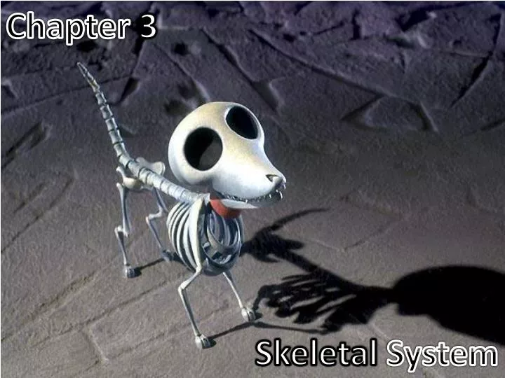
Chapter 3. Skeletal System. The Framework. Support – acts as an internal ‘scaffold’ upon which the body is built. Locomotion – provides attachment for muscles, which operate a system of levers, i.e. the bones, to bring about movement

E N D
Chapter3 SkeletalSystem
The Framework • Support – acts as an internal ‘scaffold’ upon which the body is built. • Locomotion – provides attachment for muscles, which operate a system of levers, i.e. the bones, to bring about movement • Protection – protects the underlying soft parts of the body, e.g. the brain in encased in the protective bony cranium of the skull • Storage – acts as a store for the essential minerals calcium and phosphate • Haemopoiesis – haemopoietic tissue forming the bone marrow manufactures the blood cells
Bone Structure and Function:Bone Shape • Long Bones – these are typical of the limb bones, e.g. femur, humerus, and also include bones of the metacarpus/metatarsus and phalanges; long bones have a shaft containing a medullary cavity filled with bone marrow • Flat bones – have an outer layer of compact bone with a layer of cancellous or spongy bone inside; there is no medullary cavity, e.g. flat bones of the skull, scapula and ribs. • Short bones – these have a similar structure to short bones but a less uniform shape; they lie in the midline and are unpaired, e.g. vertebrae • Irregular bones – have similar structure to short bones but a less uniform shape; they lie in the midline and are unpaired, i.e. vertebrae • Specialized types of Bone: • Sesamoid bones – these are sesame-seed-shaped bones that develop within a tendon (and occasionally a ligament) that runs over an underlying prominence; they serve to change the angel at which the tendon passes over the bone and thus reduce ‘wear and tear’, e.g. the patella associated with the stifle joint. • Pneumatic Bones – these contain air filled spaces known as sinuses that have the effect of reducing the weight of the bone, e.g. maxillary and frontal bones. • Splanchnic Bone - this is bone that develops in a soft organ and is unattached to the rest of the skeleton, e.g. the os penis (the bone within the penis of the dog and cat)
Bone Structure and Function:Development of bone • Difference between osteoblasts and osteoclasts: • Osteoblasts are the cells responsible for laying down new bone • Osteoclasts are the cells who destroy or remodel bone • The process by which bone is formed is called Ossification and there are two types: • Intramembranous ossification: this is the process by which the flat bones of the skull are formed. The osteoblasts lay down the bone between two layers of fibrous connective tissue. This is no cartilage template. • Endochondral ossification: this type of ossification involves the replacement of a hyaline cartilage model within the embryo by bone. The process starts in the developing embryo but is not completed fully until the animal has reached maturity and growth has ceased. The long bones of the limb develop by this method.
Endochondral Ossification Process: • A cartilage model develops within the embryo. • Primary centres of ossification appear in the diaphysis or shaft of the bone. The cartilage is replaced as the osteoblasts lay down bone, which gradually extends towards the ends of the bone. • Secondary centres of ossification appear in the epiphyses or ends of the bone, continuing the bone development. • Osteoclasts then start to remove bone from the centre of the diaphysis to form the marrow cavity, while the osteoblasts continue to lay down bone in the outer edges. • Between the diaphysis and epiphyses a narrow band of cartilage persists. This is the growth plate or epiphyseal plate, which allows the bone to lengthen while the animal is growing. Eventually, when the animal has reached its final size, this will be replaced by bone and growth will no longer be possible. The epiphyseal plate is then said to have ‘closed’ and the time at which it happens is different for each type of bone.
Bone disease to look out for… • Rickets: • A disease of young growing animals caused by a nutritional deficiency of vitamin D or phosphorus. The bones fail to calcify and become bowed, and the joints appear swollen because of enlargement of the epiphyses. Any animal kept permanently inside is at risk of developing this because vitamin D is formed by the action of ultraviolet light on the skin.
Bone Structure and Function:The Skeleton • Three divided parts of the skeleton: • Axial Skeleton – runs from the skull to the tip of the tail and includes the skull, mandible, and vertebrae and also the sternum. • Appendicular skeleton – the pectoral (front) and pelvic (hind) limbs and the shoulder and pelvic girdles that attach (or append) them to the body • Splanchnic skeleton – in the dog and cat, this is represented by the os penis within the tissue of the penis.
Things to know… • Tuberosity/trochanter/tubercle – protuberances on bones, which are usually for the attachment of muscles • Trochlea – bony structures through or over which tendons pass; they are usually grooves in the bone and allow tendons to act as pulleys • Condyle – a rounded projection on a bone, usually for articulation with another bone • Epicondyle – a projection of bone on the lateral edge above its condyle • Foramen – an opening or passage into or through a bone, e.g. to allow the passage of blood vessels and nerves • Fossa – a hollow or depressed area on a bone • Head, neck, and shaft are used to describe parts of a long bone • Tendon – connects muscle to bone • Ligament – connects one bone to another bone.
The Axial Skeleton:The Skull The bones of the head include the Skull, nasal chambers, mandible, or lower jaw and hyoid apparatus. The functions of the skull are: • To house and protect the brain • To house the special sense organs – eye, ear, nose, and tongue. • To house and provide attachment for parts of the digestive system – teeth and tongue, etc. • To provide attachment for the hyoid apparatus and the numerous muscles of mastication and facial expression. • To provide a bony cavity through which air can enter the body. • To communicate – the muscles of facial expression are found on the head and are an important means of communication.
The Axial Skeleton:The Skull: Nasal Chambers • The most rostral parts of the skull carries the nasal chamber, the sides of which are formed by the maxilla and the roof by the nasal bone. • The nasal chamber is divided lengthways into two by a cartilaginous plate called the nasal septum. • Each of the chambers is filled with delicate scrolls of bone called the nasal turbinates or conchae. • These are covered in ciliated mucous epithelium. • At the back of the nasal chamber, forming a boundary between the nasal and cranial cavities, is the ethmoid bone. • In the centre of this bone is the cribriform plate – a sieve-like area perforated by numerous foramina through which the olfactory nerves pass from the nasal mucosa to the olfactory bulbs of the brain. • The roof of the mouth is called the hard palate and is formed from three bones on the ventral aspect of the skull: • The incisive bone or premaxilla – the most rostral and carries the incisor teeth • Part of the maxilla • The palatine • Many of the bones of the skull are joined together by fibrous joints called sutures. • Sutures are firm and immovable joints but allow for expansion of the skull in a growing animal.
The Axial Skeleton:The Skull: Mandible • The mandible or lower jaw is comprised of two halves or dentaries, joined together at the chin by a cartilaginous joint called the mandibular symphysis. • Each half is divided into a horizontal part, the body, and a vertical part, the ramus. The body carries the sockets or alveoli or the teeth of the lower jaw. • The ramus articulates with the rest of the skull at the temporomandibular joint via a projection called the condylar process. • A rounded coronoid process, which projects from the ramus into the temporal fossa, is the point to which the temporalis muscle attaches. • There is a depression on the lateral surface of the ramus, the masseteric fossa, in which the masseter muscle lies.
The Axial Skeleton:The Skull: Cranium The caudal part of the skull that provides the bony ‘case’ in which the brain sits is called the Cranium. The bones of the cranium include: • Parietal – forms much of the dorsal and lateral walls of the cranium • Temporal – lies below the parietal bone on the caudolateral surface of the skull. The most ventral part of the temporal bone forms a rounded prominence called the Tympanic bulla, which houses the structures of the middle ear. There is an opening into the tympanic bulla, called the external acoustic meatus, which in life is closed by the tympanic membrane or eardrum. The cartilages of the external ear canal are attached to this region. • Frontal – forms the front aspect of the cranium or ‘forehead’. Contains an air-filled chamber called the frontal sinus, which connects to the nasal chamber • Occipital – this lies at the base of the skull on the caudal aspect. In this region there is a large hole called the foramen magnum, through which the spinal cord passes. On either side is a pair of bony prominences, the occipital condyles. These articulate with the first cervical vertebra or atlas. At the side of the occipital condyles are the jugular processes, which are sites for muscle attachment. • Sphenoid – this lies on the ventral aspect of the skull, forming the floor of the cranial cavity. It is penetrated by many small foramina through which nerves and blood vessels pass. • Sagittal crest – a ridge of bone on the dorsal midline surface of the skull, which can be prominent in muscular dogs • Zygomatic – the zygomatic arch is an arch of bone that projects laterally from the skull, forming the ‘cheekbone’. • Lacrimal – lies at the base of the orbit, which houses the eye, and is the region through which the tears drain from the eye into the nose.
The Axial Skeleton:The Skull: Hyoid Apparatus • The hyoid apparatus lies in the intermandibular space and consists of a number of fine bones and cartilages joined together in an arrangement that resembles a trapeze. • The hyoid apparatus is the means by which the larynx and tongue are suspended from the skull. • The Apparatus articulates with the temporal region of the skull in a cartilaginous joint.
The Axial Skeleton:The Skull: Skull Shapes • The shape of the skull varies between species. • In the domestic cat the skull is much more rounded or ‘apple-shaped’ than it is in the dog, and there is little difference between the various cat breeds. • In the dog, although the basic anatomy remains the same, the overall appearance differs greatly between the different breeds. • Three morphological forms of dog skull are recognized: • Dolichocephalic – the head particularly the nose, is long and narrow, e.g. Greyhound, borzoi, Afghan hound • Mesaticephalic – (mes meaning ‘middle’)is the ‘normal’ or average shape of the dog skull, e.g. Beagle, Labrador, Pointer • Brachycephalic – the cranium is often more rounded and the nose is short and may be pushed in, because of shortening of the nasal chambers, hard palate and mandible, e.g. Bulldog, Pekinese, Boxer, Pug.
The Axial Skeleton:The Vertebrae • The vertebral column is comprised of a number of bones arranged in a series along the midline of the body and extending from the base of the skull to the tip of the tail. • The vertebrae are divided into regions depending upon their position in the body: • Cervical – neck region • Thoracic – thoracic region • Lumbar – lower back or abdominal region • Sacral – croup or pelvic region • Caudal or coccygeal – in the tail. • Each species has a characteristic number of vertebrae within each region, which is written as a formula. • In the dog and cat this formula is: C7, T13, L7, S3, Cd20-23 • The functions of the vertebral column are: • To stiffen the body axis and help maintain posture • To enclose and protect the spinal cord • To shield and protect the softer underlying structures of the neck, thorax, abdomen, and pelvis.
The Axial Skeleton:The Vertebrae: Basic Plan Of a Vertebra • The vertebrae within all regions of the vertebral column are basically similar in structure, although each region shows slight difference related to function • A typical vertebra consists of a roughly cylindrical ventral body with a convex cranial end and a concave caudal end • This arrangement enables the bodies to fit together, creating a flexible rod • The body is topped by an arch, called the vertebral or neural arch, which forms a tunnel-like vertebral foramen through which the spinal cord passes • When linked together, the vertebral foramina constitute the spinal canal • The neural arch has a dorsal projection called the spinous process or neural spine, which varies in height and size from one region of the vertebral column to another • On either side of the vertebra there are laterally projecting processes, the transverse processes, which also vary in shape and size between regions • He transverse processes divide the muscles of the vertebral column into dorsal or epaxial and ventral or hypaxial divisions
On the cranial and caudal edges of the arch of each vertebra there are cranial and caudal articular processes • These form a synovial joint with those of the adjacent vertebrae, creating a flexile rod running in the midline of the body • There are also a number of other processes, which are sites for muscle attachment • Lying between the bodies of each pair of vertebrae is a fibrocartilaginous intervertebraldisc, which acts as a ‘shock’ absorber, preventing damage to the spinal cord • The intervertebral disc is composed of a tough fibrous connective tissue outer area, called the annulus fibrosus, and a core of gelatinous material called the nucleus pulposus • The vertebrae are linked by ligaments that run between them and they articulate with one another by two types of joint: • Cartilaginous – between the bodies of each vertebra • Synovial – between the cranial and caudal articular processes
Problems: Slipped Disc • In the case of a slipped disc, the annulus fibrosus ruptures and the pulpy centre of the disc protrudes outward • This puts pressure either on the spinal cord or on the associated nerves leaving the cord, causing the animal to show symptoms ranging from pain to paralysis due to the loss of motor function
The Axial Skeleton:The Vertebrae: Regional Variations • Cervical vertebrae– • There are always seven cervical vertebrae in the neck of all mammals • The first cervical vertebra or atlas has a unique and distinctive shape • The atlas does not have a body or a spinous process, but consists of two large wing-like lateral masses joined by a ventral and dorsal arch • The second cervical vertebra or axis is also unusual and has an elongated, blade-like spinous process, which serves as a point of attachment for neck muscles • A strong ligament, called the nuchal ligament, also attaches to the spinous process and extends from the axis to the first thoracic vertebra • On the cranial aspect of the axis, a projection of bone called the dens or odontoid process fits into the vertebral foramen of the atlas and serves as a pivot around which the atlas can be rotated • The remaining cervical vertebrae follow the basic vertebral plan, and get progressively smaller as they advance towards the junction with the thoracic vertebrae
Thoracic vertebrae – • There are usually 13 thoracic vertebrae • Their distinguishing feature is their tall spinous processes and short bodies • They articulate with the ribs at two sites: • The costal fovea: which forms a synovial joint with the head of the rib • The transverse fovea: which forms a synovial joint with the tubercle of the rib • The height of the spinous processes decreases as the series progresses towards the lumbar region
Lumbar Vertebrae – • There are usually seven lumbar vertebrae • These vertebrae have large bodies and long transverse processes angled cranioventrally, to which the lumbar muscles attach • The synovial joint between the atlas and the occipital condyles of the skull allows nodding movements of the head, and the synovial joint between the atlas and axis allows a pivotal movement so that the head can turn in all directions
The Axial Skeleton:The Vertebrae: Regional Variations • Sacral Vertebrae – • These three vertebrae are fused together to form the sacrum in the adult dog and cat • The sacrum forms a fibrosynovial joint with the wing of the ilium of the pelvic girdle: the sacroiliac joint • Caudal or Coccygeal Vertebrae – • These vary in number and shape according the length of the tail • The first few resemble the lumbar vertebrae but they get progressively smaller and simpler throughout the series • The last few caudal vertebrae are reduced to little rods of bone
The Axial Skeleton:The Vertebrae: The Ribs and Sternum • The ribs form the walls of the bony thoracic cage that protects the organs of the chest • There are 13 pairs of ribs in the dog and cat, which articulate with the thoracic vertebrae • A rib is a flat bone consisting of compact bone on the outside packed with cancellous bone on the inside • Each rib has a bony dorsal part and a cartilaginous ventral part – the costal cartilage • The most dorsal part of the bony rib has two projections: the head which articulates with the costal fovea of the vertebra and the tubercle or neck which articulates with the transverse fovea of the appropriate thoracic vertebra • During parturition, under the influence of the hormone relaxin, the sacroiliac ligament relaxes and softens so that the pelvis can stretch, enabling the fetuses to pass out through the birth canal
The costal cartilage articulates with the sternum, either directly or indirectly • The first eight pairs of ribs attach directly to the sternum and are called the sternal ribs • The ribs are called asternal or ‘false’ ribs, and they attach via their costal cartilages to the adjacent rib, forming the costal arch • The last ribs have no attachment at their cartilaginous ends, which lie free in the abdominal muscle – this pair are called the ‘floating’ ribs • The space between each successive pair f ribs is called the intercostal space and is filled by the intercostal muscles of the trunk
The sternum forms the floor of the thoracic cage and is composed of eight bones, the sternebrae, and the intersternebral cartilages • The most cranial sternebra is the manubrium, which projects in front of the first pair of ribs and forms part of the cranial thoracic inlet • Sternebrae 2-7 are short cylindrical bones • The last sternebra is longer and dorsoventrally flattened and is called the xiphoid process • Attached to the xiphoid process and projecting caudally is a flap of cartilage called the xiphoid cartilage • The linea alba attaches to this • Between each pair of sternebrae are cartilaginous discs called the intersternebral cartilages
The Appendicular Skeleton • The Appendicular skeleton is composed of the pectoral (or fore) limb and the pelvic (or hind) limb and the shoulder and pelvic girdles that attach these to the body • The forelimb has no bony connection to the trunk, only being attached by muscles • This absorbs the ‘shock’ at the point when the limb takes the animal’s weight in four-legged animals or running quadrupeds • This differs from primates, which generally walk on their hind legs and so have evolves a pectoral girdle with a clavicle • However, the hindlimb does have a bony articulation in the pelvic girdle, which forms the platform for the muscles that provide the propulsive force as the animal is running
The Appendicular Skeleton:Bones on the Forelimb • Clavicle – frequently absent in the dog. When present, it is just a remnant of bone that lies in the muscles cranial to the shoulder joint – is it described as being vestigial. The clavicle is normally present in the cat but does not articulate with other bones. • Scapula – also called the shoulder blade. It is a large, flat bone found on the lateral surface of the trunk at the junction of the neck and ribs. It has a prominent ridge or spine running down the middle of its lateral surface. This divides the lateral surface into two regions: the supraspinous fossa and infraspinous fossa. On the distal end of the spine these is a bony projection called the acromion. At the distal end of the scapula the bone narrows at the neck and there is a shallow articular socket, called the glenoid cavity, which forms the shoulder joint with the head of the humerus. The medial surface of the scapula is flat and comparatively smooth.
Humerus – this is the long bone forming the upper forelimb. It articulates proximally with the scapula at the shoulder joint, and distally with the radius and ulna at the elbow joint. The proximal end of the humerus consists of a large rounded projection, the head. Cranial and lateral to the head these is a large prominence, called the greater tubercle. Another prominence, the lesser tubercle, lies medial to the head. Both of these are sites for attachment of the muscles that support the shoulder joint. Distal to the head is the neck, attached to the slightly twisted shaft of the bone. On the distal end of the humerus are the medial and lateral epicondyles, between which is the condyle. Just proximal to this is a deep hollow called the olecranon fossa. This receives the anconeal process of the ulna. There is also a hole in the centre of the condyle called the supratrochlear foramen. N.B. There is no supratrochlear foramen in the cat.
Radius and Ulna – These are both long bones that lie side by side in the forearm. At the proximal end of the ulna is a projection known as the olecranon, which forms the point of the elbow in the front of this is a crescent shaped concavity called the trochlear notch, which articulates with the distal humerus. At the top of the trochlear notch is a beak-like projection called the anconeal process, which sits within the olecranon fossa of the humerus when the elbow is extended. Distally, the ulna narrows to a point called the lateral syloid process. The radius is a rod-like bone, shorter than the ulna. At the proximal end is a depression, the fovea capitis, which articulates with the humerus. At the distal end of the radius there is a pointed projection called the medial styloid process.
Carpus – this is composed of seven short bones, the carpal bones, arranged in two rows. The proximal row has three bones, the most medial being the radial carpal bone, which articulates proximally with the radius. The ulnar carpal bone articulates proximally with the ulna. The accessory carpal bone lies on he lateral edge and projects caudally. Distally, the first row of carpal bones articulates with the second row of four carpal bones. The carpal bones also articulate with each other within the row. • Metacarpus – this is composed of five small long bones. In the dog and cat the first metacarpal bone, i.e. the most medial, is much smaller than the other metacarpal bones, and is non-weight bearing. This forms part of dew claw. The metacarpals articulate proximally with the distal row of carpal bones and distally with the phalanges.
Digits – these are composed of the phalanges, which are long bones. Each digit has three phalanges, except digit I – the dew claw – which has only two. The proximal phalanx articulates with a metacarpal bone. The middle phalanx articulates with the phalanx above and below it. The distal phalanx ends in the ungual process, which forms part of the claw. • There are pairs of small sesamaoid bones behind the metacarpophalangeal joints and the distal joints between the phalangeal bones.
The Appendicular Skeleton:Bones on the Hindlimb • Pelvis – this is the means by which the hindlimb connects to the body. It consists of two hip bones or ossa coxarum, which join together at the public symphysis. They form a firm articulation with the sacrum at the sacroiliacjoint. Each hip bone is formed from three bones – the ischium, ilium, and pubis – grouped around one very small bone called the acetabular bone. The largest of these bones is the ilium, which has a broad cranial expansion called the wing. The ischium has a prominent caudal projection called the ischial tuberosity. The ilium, ischium, and pubis meet each other at the acetabulum, which is the articular socket in which the head of the femur sits, forming the hip joint. The hip joint is a ball-and-socket joint. • Hip dysplasia is an inherited condition that affects a range of larger breeds of dog, e.g. Labradors, Retrievers, German Shepherds. It is caused by malformation of the femoral head and/or a shallow or malformed acetabulum, resulting in subluxation of the hip joint leading to osteoarthritis. There is a BVA/KC scheme to help identify affected dogs and to advise breeders on the choice of breeding stock.
The head of the femur is held in place by a ligament known as the teres or round ligament, which attaches to a non-articular area within the joint cavity called the acetabular fossa. On either side of the pubic symphysis is a large hole called the obturator foramen that serves to reduce the weight of the pelvic girdle and to provide extra surface area for the attachment of muscles and ligaments. • Femur – this is a long bone and forms the thigh. On the proximal femur the articular head faces medially to articulate with the acetabulum of the pelvis. The head is joined to the shaft of the neck. Lateral to the head is a projection called the greater trochanter and on the medial side is another smaller projection called the lesser condyle, which articulate with the tibia at the stifle joint. The patella runs between these condyles in the trochlea groove. • The patella is a sesamoid bone found within the tendon of insertion of the quadriceps femoris muscle, which is the main extensor of the stifle. Two more sesamoid bones, called the fabellae are found behind the stifle in the origin or the gastrocneius muscle. They articulate with the condyles of the femur.
Tibia and Fibula – these long bones form the lower led. The tibia and fibula lie parallel to each other, the more medial bone, the tibia, being the much larger of the two. The tibia is expanded proximally where it articulates with the femur. On the dorsal surface there is a prominence called the tibial crest for attachment of the quadriceps femoris muscle. Distally, the tibia has a prominent protrusion, the medial maleolus, which can be palpated on the medial aspect of the hock. The fibula is a thin long bone lying laterally to the tibia. It ends in a bony point called the lateral malleolus. • Tarsus – this is formed from seven short bones, the tarsel bones, arranged in three rows. The two bones forming the proximal row, the talus and calcanus, articulate with the distal end of the tibia and fibula at the hock joint. The talus, or tibial tarsal bone, is the most medial and has a proximal trochlea, which is shaped to fit the end of the tibia. The calcaneus, or fibular tarsal bone, is positioned laterally and has a large caudal projection known as the tuber calcis, which forms the ‘point’ of the hock. • In some small breeds of dogs, e.g. Yorkshire Terrier, the patella may slip out of place, causing extreme pain and difficulty in extending the stifle join. This is an inherited condition and is due to mal positioning of the tibial crest or too shallow a trochlear groove on the distal end of the femur.
Metatarsus and Digits - these closely resemble the pattern of the metacarpus and digits in the forepaw. The metatarsus is composed of four metatarsal bones, although some breeds possess five, having a small metatarsal I or hind dew claw.
The Splanchnic Skeleton • This is composed of the splanchnic bones. • A splanchnic bone is a bone that develops in soft tissue and is unattached to the rest of the skeleton. • The only example of a splanchnic bone in the dog and cat is the bone of the penis, the os penis. • The urethrea lies in the urethral groove, on the ventral surface of the os penis in the dog. • In the cat the urethral groove is on the dorsal surface of the os penis, because of the different orientation of the penis. • The cow has a splanchnic bone in its heart, called the os cordis, while birds has splanchnic bones forming a rim around the eye to provide strength to the large eyeball.
The Splanchnic Skeleton:Joints: Fibrous Joints • Fibrous joints are immovable joints and the bones forming them are united by dense fibrous connective tissue, e.g. in the skull fibrous joints unite the majority of the component bones and are called sutures. • The teeth are attached to the bony sockets in the jaw bone by fibrous joints. • Fibrous joints are also classed as synarthroses, i.e. a type of joint that permits little or no movement. • Some cartilaginous joints also fall into this category.
The Splanchnic Skeleton:Joints: Cartilaginous joints • Cartilaginous joints allow limited movement or no movement at all and are united by cartilage, e.g. the pubic symphysis connecting the two hip bones and the mandibular symphysis joining the two halves of the mandible. • Both these joints are also classed as synarthroses. • Some cartilaginous joint may also be classed as amphiarthroses, which allowed some degree of movement between the bones, e.g. between the bodies of the vertebrae allowing for limited flexibility of the spinal column.
Synovial Joints • Synovial Joints or diahroses allow a wide range of movement. • In synovial joints, the bones are separated by a space filled with synovial fluid known as the joint cavity. • A joint capsule surrounds the whole joint; the outer layer consists of fibrous tissue, which serves as protection, and the joint cavity is lined by the synovial membrane, which secretes synovial fluid. • This lubricates the joint and provides nutrition for the hyaline articular cartilage covering the ends of the bone • Synovial fluid is a straw colored viscous fluid that may be present in quite large quantities in large joints, especially in animals that have a lot of exercise.
Some synovial joints may have additional stabilisation from thickened ligaments within the fibres of the joint capsule • These are most commonly found on either side of the joint, where they are called collateral ligaments • However, other synovial joints have stabilising ligaments attached to the articulating bones within the joint – these are known as intracapsular ligaments and examples include the cruciate ligaments within the stifle joint. • A few synovial joints possess one or more intraarticular fibrocartilaginous discs or menisci within the joint cavity. • These are found in the stifle joint – it has two crescent shaped menisci – and in the temporomandibular joint between the mandible and he skull • These structures help to increase the range of movement of the joint and act as ‘shock absorbers’, reducing wear and tear
Synovial joints allow considerable freedom of movement between the articulating bones, the extent of which depends upon the type of synovial joint • The movement allowed by a synovial joint may be in a single plane only, or in multiple planes • Synovial joints can be further classified into subcategories based upon the types of movement that they allow.
The Range Of Movements That Are Possible In Synovial Joints: • Flexion/extension – these are antagonistic movements of a joint • Flexion reduces the angle between two bones, i.e. bends the limb • Extension increases the angle between two bones, i.e. straightens the limb • Abduction/adduction – these movements affect the whole limb; • Abduction (means to ‘take away’) moves a body part away from the median plane or axis, e.g. moving the leg out sideways • Adduction moves a body part back towards the central line or axis of the body, e.g. moving the leg back to standing position • Rotation – the moving body part ‘twists’ on its own axis, i.e. it rotates either inwardly or outwardly • Circumduction – the movement of an extremity, i.e. one end of a bone, in a circular pattern. • Gliding/sliding – the articular surfaces of the joint slide over one another • Protraction – the animal moves its limb cranially, i.e. advances the limb forward, as when walking • Retraction – the animal moves the limb back towards the body
Types of Synovial Joints • Plane/gliding – allows sliding of one bony surface over the other, i.e. joints between the rows of carpal and tarsal bones • Hinge – Allows movement in one plane only, i.e. elbow; stifle • Pivot – consists of a peg sitting within a ring; allows rotation, i.e. atlantoaxial joint • Condylar – consists of a convex surface (condyles) that sits in a corresponding concave surface; allows movement in two planes (flexion, extension, and overextension), i.e. hock or (tarsus) • Ball and Socket – consists of a rounded end or ball, sitting within a socket or cup; allows a great range movement