1 / 48

Cell Junctions and extra cellular matrix II

chapter 19. Cell Junctions and extra cellular matrix II. Desmosomes and hemidesmosomes both link to intermediate filaments. Cadherins also bind intermediate filaments together. Integrins link extracellular matrix to cytoskeleton.

mariah
Download Presentation

Cell Junctions and extra cellular matrix II

An Image/Link below is provided (as is) to download presentation Download Policy: Content on the Website is provided to you AS IS for your information and personal use and may not be sold / licensed / shared on other websites without getting consent from its author. Content is provided to you AS IS for your information and personal use only. Download presentation by click this link. While downloading, if for some reason you are not able to download a presentation, the publisher may have deleted the file from their server. During download, if you can't get a presentation, the file might be deleted by the publisher.

E N D

Presentation Transcript


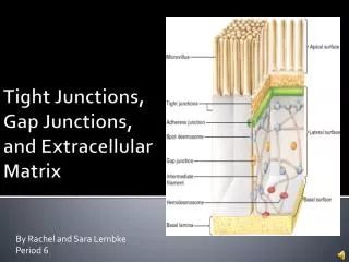
  1. chapter 19 Cell Junctions and extra cellular matrix II

  2. Desmosomes and hemidesmosomes both link to intermediate filaments

  3. Cadherins also bind intermediate filaments together

  4. Integrins link extracellular matrix to cytoskeleton

  5. Size of gap junction channel can be determined with fluorescent molecules of different sizes

  6. A Gap junction connexon is made up of 6 transmembrane connexin subunits

  7. Images of gap junctions

  8. The permeability of gap junctions can be regulated dopamine treatment reduces permeability of gap junctions to a fluorescent dye

  9. Summary of cell junctions found in the cell

  10. Summary of cell junctions found in the cell

  11. In plant cells plasmodesmata perform many of the same functions as gap junctions

  12. View of plasmodesmata

  13. Structure of Cadherin – responsible for cell to cell adhesion in vertebrate cells

  14. Ca2+ is required to establish cell to cell connection through cadherin

  15. Three mechanisms that mediate cell-cell adhesion cadherins least common

  16. Cadherins allow cells to sort themselves

  17. Catenins join cadherins to actin filaments in adherence junctions

  18. Summary of junctional and non-junctional adhesive mechanisms

  19. Cells surrounded by extracellular matrix

  20. Cross sectional view of intestinal wall

  21. Connective tissue underlying the epithelium contains a variety of cells and extracellular matrix components

  22. Fibroblasts surrounded by collagen fibrils

  23. Glycosaminoglycan (GAG) chains occupy large amounts of space and form hydrated gels

  24. Hyaluronan, a simple GAG, occupies a large amount of space, thereby providing mechanical support

  25. Repeating disaccharide unit of hyaluronan

  26. GAG and its core protein form proteoglycan

  27. Comparison of proteoglycans with a ribonuclease

  28. picture of aggrecan aggregate, a large proteoglycan

  29. structure of Aggrecan aggregate

  30. Collagen fibril embedded in proteoglycan molecules

  31. Fibroblast surrounded by collagen fibrils

  32. Collagen fibrils in tadpole skin This arrangement is also found in bone and the cornea

  33. Elastic fibers in a dog’s aorta

  34. Each elastin protein in the network can expand and contract as a coil

  35. Fibronectin is an extracellular protein that helps cells attach to the matrix

  36. Connective tissue underlying the epithelium contains a variety of cells and extracellular matrix components

  37. basal lamina in the cornea of a chick’s embryo

  38. A model of the basal lamina

  39. Cross Section of Kidney

  40. Filtration is one step in urine formation

  41. Structure of Nephrons

  42. Inside Bowman’s Capsule

  43. Basal Lamina plays an important role in filtration

More Related