MATRIX/CELL-CELL/CYTOSKELETAL TENSION
200 likes | 388 Views
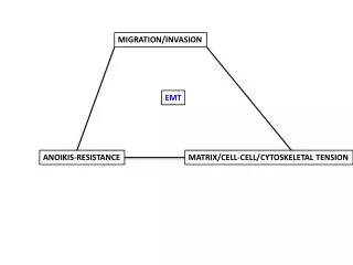
MIGRATION/INVASION. EMT. ANOIKIS-RESISTANCE. MATRIX/CELL-CELL/CYTOSKELETAL TENSION. Mina Bissell. “DYNAMIC RECIPROCITY”. Changes in tensional forces (cell-cell and/or cell-matrix can affect EMT-related cell behavior. Regulation of Epithelial- Mesenchymal Transition

MATRIX/CELL-CELL/CYTOSKELETAL TENSION
E N D
Presentation Transcript
MIGRATION/INVASION EMT ANOIKIS-RESISTANCE MATRIX/CELL-CELL/CYTOSKELETAL TENSION
Mina Bissell “DYNAMIC RECIPROCITY”
Changes in tensional forces (cell-cell and/or cell-matrix can affect EMT-related cell behavior
Regulation of Epithelial-Mesenchymal Transition by Transmission of Mechanical Stress through Epithelial Tissues NikolceGjorevski & ElineBoghaert & Celeste M. Nelson Cancer Microenvironment DOI 10.1007/s12307-011-0076-5 Fig. 3 Endogenous mechanical stress patterns EMT. a In monolayers of epithelial cells, stress is concentrated at the free edges and corners of the tissues. b Under these conditions, MRTF-A accumulatesin the nuclei of cells located at the free edges and corners of the tissue, but is sequestered in the cytoplasm of cells located in the center of the tissue. c When treated with TGFβ, cells located in high stress regions undergo EMT
Regulation of Epithelial-Mesenchymal Transition by Transmission of Mechanical Stress through Epithelial Tissues NikolceGjorevski & ElineBoghaert & Celeste M. Nelson Cancer Microenvironment DOI 10.1007/s12307-011-0076-5 Fig. 2 Regulation of MRTF-A by mechanical stress. a Increased mechanical stress causes increased actin polymerization, thereby decreasing the cytoplasmic pool of G-actin and increasing the nuclear localization of MRTF-A by triggering its dissociation from G-actin. b Under conditions of low mechanical stress a larger cytoplasmic pool of G-actin sequesters MRTF-A in the cytoplasm
Mechanisms by Which the Extracellular Matrix and Integrin Signaling Act to Regulate the Switch Between Tumor Suppression and Tumor Promotion Patricia J. Keely Journal of Mammary Gland Biology and Neoplasia Fig. 3 Integrin-mediated signaling pathways regulate normal epithelial polarity and differentiation. Adhesion of α6β4 integrin to the basal lamina (B. Lamina) results in activation of FAK, and subsequent signaling to Rac to promote cell proliferation and survival via p21 and NFkB. Signaling through Rac to promote basal secretion of laminin-5 (LN-5) helps establish cell polarity. Adhesion to the basal lamina prevents apoptosis via PAK activation of Bcl2 and Bad. Loss of adhesion to the basal lamina results in apoptosis, necessary for luminal clearing. In the case of tumorigenesis, tumor cells degrade the basal lamina, and invade into the stromal matrix. Tumor cells can secrete their own autocrine LN-5 and prevent apoptosis. Moreover, α2β1 integrin adhesion to collagen promotes cell proliferation and survival via activation of FAK, pERK, and PI-3-K pathways. Activated pFAK antagonizes p53 to prevent apoptosis
Matrix density-induced mechanoregulation of breast cell phenotype, signaling, and gene expression through a FAK-ERK linkage Paolo P. Provenzano,1,2,3,4* David R. Inman,1,4 Kevin W. Eliceiri,2,3 and Patricia J. Keely1,2,3,4* Oncogene. 2009 December 10; 28(49): 4326–4343.
Mechanical tugging force regulates the size of cell–cell junctions Zhijun Liua,1, John L. Tanb,1, Daniel M. Cohena,1, Michael T. Yanga, Nathan J. Sniadeckia,SamiAlomRuiza, Celeste M. Nelson, and Christopher S. Chen 9944–9949 ∣ PNAS ∣ June 1, 2010 ∣ vol. 107 ∣
CAN THESE FACTORS AFFECT EMT VIA CYTOSKELETON: A.CORTACTIN (WEED) B. MYOSIN ISOFORMS (WYSOLMERSKI) C. FAK (SCHALLER) D. ANKYRIN-G (FRISCH)
2. EMT can affect cell-matrix and cell-cell tension, with important cellular responses
Figure 3. Deflection images of alveolar epithelial cells measured by atomic force microscopy. A549 cells were grown on chamber slides and treated with TGF-β1 (5 ng/ml) for 48 h. Cells were then fixed using a mixture of paraformaldehyde and glutaraldehyde. Images were acquired using a cantilever with 2.8 N/m spring constant in PBS buffer at room temperature. (A-D)Visualization of the cell ultrastructure revealed numerous distinct filamentous structures (arrows) in those cells treated with TGF-β1 on glass (B) or on collagen I (D), representative of F-actin stress fibers. In contrast, untreated cells (A,C) exhibited indistinct filament arrangements. Images shown are representative data of 3 independent experiments. Bars 10 μm.
Approaches for investigating intercellular forces and mechanosensing through cell surface adhesion proteins. (a) In traction force microscopy, fluorescent particles embedded in soft gels serve as fiduciary markers to quantify the traction force exerted by attached cells. Bead displacement maps are converted to local traction forces, which are represented in heat maps (b), which indicate cell traction force distributions. Here, two cells are in contact, and the cell boundary is indicated in (a) and (b) (from [27]). (c) Micro-arrays of elastomeric pillars are used to determine cell tractions from the cell-induced deflections of the pillars. Figures c and d show two adhering cells on the array. (d) Between cell doublets constrained to a bowtie pattern on the array, increases in the net force at the cell–cell contact correlated with an increase in the size of the intercellular junction, indicated by bcatenin (green) (from [29]). (e) In magnetic twisting cytometry (MTC), cadherin-modified beads are attached to the cell surfaces. (f) The magnetized beads are subject to an orthogonal, oscillating magnetic field, H, which induces a torque, T, on the bead. The amplitude of the resulting bead displacement, D, reflects the stiffness of the bead-cell-cytoskeletal linkage. Current Opinion in Cell Biology 2011, 23:523–530 www.sciencedirect.com
Transcriptional crosstalk between TGF-β and stem cell pathways in tumor cell invasion Jonas Fuxe,1,* Theresa Vincent2 and Antonio Garcia de Herreros Cell Cycle 9:12, 2363-2374; June 15, 2010 Invasion genes (?) Anoikis-resistance genes (?)
Targets of miR-200c mediate suppression of cell motility and anoikis resistance Erin N Howe, Dawn R Cochrane and Jennifer K Richer Howe et al. Breast Cancer Research 2011, 13:R45 http://breast-cancer-research.com/content/13/2/R45 They guessed Moesin Was important from microarray. Turned out to be right. But a functional screen would be more powerful!
Cancer Metastasis Rev. 2009 Jun;28(1-2):15-33. EMT, the cytoskeleton, and cancer cell invasion. Yilmaz M, Christofori G.
Cancer Cell 19, 372–386, March 15, 2011 TwistPDGRinvadopodiainvasion Other stuff
HOW DOES EMT TRANSLATE INTO INCREASED MIGRATION/INVASION? --HOW DOES EMT ALTER CELL-CELL OR CELL-MATRIX ADHESIONS AND THE TENSION THEY PRODUCE? (LEGLEITER, WYSOLMERSKI, SCHALLER) --DO SPECIFIC EMT-REGULATORY GENES (RUPPERT—KLF4, GLI, TGF-B; FRISCH—ZEB1, GRHL2; CTBP, TGF-B; IVANOV—ZEB1) AFFECT CELL STRUCTURES (WYSOLMERSKI, LEGLEITER—CYTOSKELETON; SCHALLER—FOCAL ADHESIONS; WEED—INVADOPODIA,LAMELLIPODIA; FRISCH-CELL JUNCTIONS) OR MAYBE EVEN OUR FAVORITE PROTEINS WITHIN THESE STRUCTURES: (WEED—SRC, ABL, CORTACTIN; WYSOLMERSKI—MYOSIN; SCHALLER—FAK; FRISCH--ANKYRIN)
WE CAN FIND OUT THROUGH COLLABORATIONS …AND PUBLISH SOME INTERESTING PAPERS (THE FIRST STEP TOWARD FUNDING)
Tensional homeostasis and the malignant phenotype. Paszek MJ, Zahir N, Johnson KR, Lakins JN, Rozenberg GI, Gefen A, Reinhart-King CA, Margulies SS, Dembo M, Boettiger D, Hammer DA, Weaver VM. Cancer Cell. 2005 Sep;8(3):241-54. Nature Reviews Molecular Cell Biology 12, 308-319 (May 2011) | doi:10.1038/nrm3112 Balancing forces: architectural control of mechanotransduction Christopher C. DuFort1, Matthew J. Paszek1 & Valerie M. Weaver J Mammary Gland BiolNeoplasia. 2011 Sep;16(3):205-19. Epub 2011 Aug 7. Mechanisms by which the extracellular matrix and integrin signaling act to regulate the switch between tumor suppression and tumor promotion. Keely PJ. Mechanotransduction at cadherin-mediated adhesions Deborah E Leckband1, Quint le Duc2, Ning Wang3 and Johan de Rooij Current Opinion in Cell Biology 2011, 23:523–530 Cancer Metastasis Rev. 2009 Jun;28(1-2):15-33. EMT, the cytoskeleton, and cancer cell invasion. Yilmaz M, Christofori G. Growth control by intracellular tension and extracellular stiffness. Assoian RK, Klein EA. Trends Cell Biol. 2008 Jul;18(7):347-52. Epub 2008 May 29. Review.