Download
1 / 58
630 likes | 836 Views
Adhesion Cell junctions. Dr. habil. Kőhidai László Assoc. Professor Dept. Genetics, Cell- and Immunobiology Semelweis University. 20 18 – ED. Significance of cell-adhesion: formation of cell la yers. Significance of cell-adh es ion: Formation of multiple layers of cells.

E N D
Adhesion Cell junctions Dr. habil. Kőhidai László Assoc. Professor Dept. Genetics, Cell- and Immunobiology Semelweis University 2018– ED
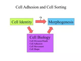
Significance of cell-adhesion: Formation of multiple layers of cells
Lymphocytes cross the endothel „Rolling” Entry Adhesion Migration homing receptor vascular addressin
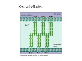
! Main adhesion molecules expressed on the cells during adhesion • Cadhedrins homophil • Selectins heterophil • Integrins heterophil • Adh. mol. with immunoglobulin homophil domain { Ca2+
! i.e. cadherin cyto- skele- ton i.c. linker prot. i.e. integrin ECM
! Cadherin Ca2+ Ca2+ • Homophil connection • Cell-cell • Ca2+-dependent binding • I.c. anchored to actin or • intermedier filamentum • Significant role in • development • of vertebrates Ca2+ catenin • Tissue-specificity: • N - nerve • P - placenta • E - epithelial actin or IF
! CAM = Cell Adhesion Molecules • 5 pcs. Ig-type domain • Ca 2+ indep. adhesion • Homophi connection - typical • Heterophil connection - rare • More than 20 variations • Expressed in the critical phase of • development Ig-like domains • Tissue-specificity: • N-CAM - neuron • L-CAM - liver In melanoma – relation between ICAM-1 density and the Metastatic activity of the tumour
! Selectins Lectin type proteins, Carbohydrate specificity Tissue-specificity: E - epithelial L - lymphoid P - placenta L selectin: it has role in the initial phase of adhesion; in newborns the level of L sel. is low – the low number of inflammations (?!)
! Integrin b • Ca2+-dependent binding • Heterophil connection • Focal contacts • Its i.c. linker proteins are • i.e.. talin, a-actinin, vinculin • RGD sequ. Is significant in • e.c. binding • Partner molecules: • fibronectin • laminin • collagen a S S Deficiency(b) – the adhesion of leukocytes affected, results the increase of inflammations
! Junctional complexes • Tight junction • Zonula adherens • Desmosome • Gap junction • Hemidesmosome • Interdigitation
Intercellular space 0.6 mm Transmembrane proteins occludin Lipid bilayer Cytoplasmic face Tight junction = zonula occludens
Negative staining to detect „insulating” effect of tight junction
! Role of tight junction • Mechanical connection • Barrier • Inhibits: • entry of water-soluble molecules • lateral diffusion of membrane-components • Ca2+ is required for the connections of occludins • Permeability: inorganic small substances • amino acids • monosaccharides
Septate junction • on the apical part of the cells • they form a continous belt (like z. adherens) • promote to stick together the cells • actin is anchored in this junction
Desmosome = macula adherens Cell to cell, snap-like junction
! cytoplasmatic plaque desmoplaquin, plactoglobin Desmosome actin intermedier filaments desmosomal cadherins i.e. desmoglein, desmocollin
TEM structure of desmosome Cytoplasmatic plaque Intermedier filaments (i.e. keratin) cadherins
! Gap junction (1958, 1968) • Bridges the 2-4 nm gap between the neighbour cells • Hexagonal channel is formed (connexon) • The wall of the channel is composed by 6 connexins • (4 helical components) • The distribution of channels is tissue- or cell-specific • due to the difference of connexins • Accross channels substances are transmitted • (max MW. 1000) by a regulated mechanism
! Role of gap junction • Transportofneurotransmitters, cAMP, • Ca 2+ • Signaling in early embryo
Plasmodesmata sER cytoplasm desmotubule cellwall membrane plasmodesmata Gap junction-like function Viral infection results in the increase of pore diameter
Plasmodesmata ER membrane desmotubule
! Hemidesmosome Fixing of epithelial cells to the basal membrane
! Basal striation
! Interdigitation Characteristic between epithelial cells
Structure of focal contact actin filament a actinin vinculin paxillin talin integrin fibronectin
Focal contacts actin vinculin
Extracellular matrix - Function • Influence on migration of the cells • Regulation of activity of molecules released • Co-receptors
! Extracellular matrix – Building blocks • Glycosaminoglycans (GAG-s) • heparan sulfate - basal membr., cell surface • keratan sulfate - cornea, bone • chondroitin sulfate – cartilage, bone, heart • hyaluronate - synovial fluid • dermatan sulfate - blood vessels, heart • „Core-proteins” aggrecan – decorin • collagen – elastin - structure • fibronectin – laminin - adhesion
Fibronectins collagen cell RGD Dimer composed by different channels – alternative splicing heparin
! Fibronectins Gly Arg Asp • Glycoproteins • Dimer-structure • RGD (Arg-Gly-Asp) • Plasma fibronectins • Gene „K.O.” experiments
Basal lamina Cell Connective tissue Layers: lamina lucida lamina densa lamina fibroreticularis Components: IV. collagen laminin perlecan entactin
SEM image of basal lamina epithelial cells basal membrane collagen fibrils
! Main proteins composing the basal lamina
! Collagen IV Entactin Perlecan Laminin
Collagen • Fibrils(diam.10-300 nm) • More than 15 types • I., II., III., V., XI. – formation of fibrils • IV., VII. – network • IX., XII. – association of fibrils
Synthesis and association of collagen filament synthesis of a-chain Pro, Lys hydroxilation glycosylation assembly organization into a fiber formation of triple helix secretion splitting of procollagen building into fibrill
Negative staining of collagen fibrils gaps between collagen molecules collagen
! B2 chain B1 chain A chain a-helix Globular domains Laminin • each chain has more types • 18 isoforms of laminin • diversities in tissues
! strech relax Elastin fibre of elastin cross-links