Chapter 9: Tools for Analyzing Gene Expression
Chapter 9: Tools for Analyzing Gene Expression. In the post-genomic era, researchers need a tool that enables the direct visualization of biological functions and GFP has turned out to be that tool. Atsushi Miyawaki, Cell 135 (2008), p. 987. 9.1 Introduction.


Chapter 9: Tools for Analyzing Gene Expression
E N D
Presentation Transcript
Chapter 9: Tools for Analyzing Gene Expression
In the post-genomic era, researchers need a tool that enables the direct visualization of biological functions and GFP has turned out to be that tool. Atsushi Miyawaki, Cell 135 (2008), p. 987
After a new gene is cloned, the next steps are to determine: • The structure of the gene. • How its expression is regulated. • The biological functions of the encoded gene product.
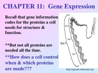
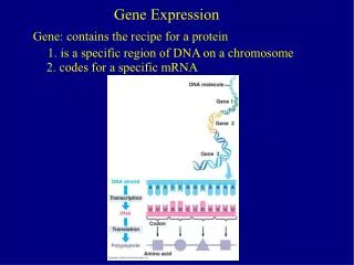
Gene expression is the production of a functional protein or RNA from the genetic information encoded in the genes. • The term encompasses both transcription and translation. • Often, gene expression is used to refer to the process of transcription only.
Overview of tools for analyzing gene regulation and function. • Resource for when tools become relevant for understanding experiments referred to in subsequent or previous chapters.
Model organisms • Each model organism is distinctively suited, as a simplified model, to the study of particular complex aspects of biology.
General attributes of model organisms • Relatively cheap and plentiful. • Inexpensive to house. • Straightforward to propagate. • Short gestation periods that produce large numbers of offspring. • Easy to manipulate in the lab. • Some have a fairly small and relatively uncomplicated genome.
Classic model organisms for molecular biology • Bacteriophage lambda () • The bacterium Escherichia coli
Some widely used eukaryotic model organisms Slime mold: Dictyostelium discoideum Ciliate: Tetrahymena thermophila Yeast: Saccharomyces cerevisiae and Schizosaccharomyces pombe Worm: Caenorhabditis elegans
Fly: Drosophila melanogaster Fish: Danio rerio Plant: Arabidopsis thaliana Mouse: Mus musculus Frog: Xenopus laevis and Xenopus tropicalis
Transfection: the introduction of DNA into eukaryotic cells. • Plasmid DNA remains extrachromosomal. • Plasmid DNA is not replicated in mammalian cells and is eventually lost by degradation and by dilution as cells divide.
Transient transfection: the introduction of DNA into cells for a short duration. • Stable transfection: Cells that have stably integrated the plasmid into a chromosome are selected for by drug resistance.
A reporter gene is a known gene whose RNA or protein levels can be measured easily and accurately. • Often used to replace other coding regions whose protein products are difficult to measure quantitatively.
Some applications of reporter genes: • The activity of the regulatory regions from another gene in different tissues or developmental stages. • The efficiency of gene delivery systems. • The intracellular fate of a gene product. • Protein-protein interactions. • DNA-protein interactions. • The success of molecular cloning efforts.
Commonly used reporter genes • Generally code for proteins with enzymatic activities or fluorescent properties not typically found in the cells of most eukaryotes. • The choice of reporter gene depends on the cell system being used, the sensitivity required, and the desired method of analysis.
CAT reporter gene assay • Chloramphenical acetyltransferase (CAT) catalyzes the acetylation of chloramphenicol, with acetyl group donated by acetyl CoA. • Acetylated chloramphenicol can be monitored by: • Autoradiography following thin-layer chromatography • Enzyme-linked immunosorbent assay (ELISA)
Analysis of gene expression Example: • Activation of reporter gene expression by overexpression of a transcription factor using a cotransfection assay.
Purification and detection tags: fusion proteins • Reporter genes can be attached to other sequences so that the reporter protein is synthesized fused to another protein. • Often a short peptide sequence that serves as an affinity or epitope tag (antigenic determinant) is used.
Fusion proteins are used for studies of: • Protein localization. • DNA-protein interactions. • Protein-protein interactions. • To make large quantities of protein for structural studies.
Commonly used purification and detection tags Protein or peptide affinity tags: • Histidine (His) tag:6-histidine • GST tag:glutathione-S-transferase
Immunotags: • c-Myc: a transcription factor • FLAG: Asp-Tyr-Lys-Asp-Asp-Asp-Asp-Lys • HA: influenza A virus haemagglutinin
Fluorescent protein tags Green fluorescent protein • Originally isolated from the jellyfish Aequorea victoria. • The fluorescence of GFP can be detected directly in living cells. • GFP can artificially be expressed effectively in every cell type and organism tested so far.
Properties of green fluorescent protein • GFP fluorophore is buried in the center of a cylinder formed by an 11-stranded -barrel. • A fluorophore is a group of atoms in a molecule responsible for absorbing light energy and producing the color of the compound. • GFP fluorophore arises from an autocatalytic post-translational modification of GFP.
Fluorescent proteins with different spectra • Mutant forms of GFP • Enhanced GFP (EGFP): Red-shifted variant • Yellow fluorescent protein (YFP) • Cyan fluorescent protein (CFP) • Red fluorescent protein from a tropical coral, Discosoma striata (RFP or DsRed) • Variants of DsRed: fruit fluorescent proteins • mCherry, pmBanana, tdTomato, etc.
Examples of use of fluorescent fusion proteins • Tracking the intracellular localization of a protein of interest. • Multiple labeling of different organelles or structures within the same cells or different tissues of cells in the same organism.
Production of recombinant protein • Over-expression of recombinant proteins in bacteria. • Over-expression of recombinant proteins in eukaryotic cells. • In vitro translation of recombinant proteins.
Fluorescence, confocal, and multiphoton microscopy • Imaging of either fixed or living tissues that have been labeled with one or more fluorescent probes.
When samples thicker than 2 m are imaged using conventional fluorescence microscopy, resolution is poor due to out-of-focus fluorescence. • Confocal and multiphoton microscopy have enabled the imaging of discrete regions of tissues at high resolution.
Fluorescence terminology • A fluorochrome is a natural or synthetic dye or molecule that can exhibit fluorescence. e.g. fluorescein isothiocyanate (FITC) • A fluorophore is a group of atoms in a molecule responsible for absorbing light energy and producing the color of the compound. • These words tend to get used interchangeably in the scientific literature.
Confocal microscopy • IIlumination is achieved by scanning one or more focused beams of light from a laser across the specimen. • IIluminated light is focused to a diffraction-limited spot. • The signal photons are focused onto a detector pinhole that rejects scattered and out-of-focus light.
By collecting a series of “optical sections” (Z series) researchers can create, with the help of sophisticated computer algorithms, high-resolution, three-dimensional images of a sample.
Multiphoton microscopy • Also known as two-photon microscopy. • The sensitivity of detection is much higher than for confocal microscopy. • Multiphoton excitation is limited to the plane of focus, thus reducing photobleaching and photodamage of samples. • Particularly useful for live cell analysis in thick tissues.
Three main types of in vitro mutagenesis • Deletion mutagenesis by PCR removes segments of DNA from a gene clone. • Linker scanning mutagenesis is the systematic replacement of each part of a gene clone to determine its function. • Site-directed mutagenesis is the introduction of specific base substitutions or small insertions at defined sites in a cloned DNA molecule.
9.5 Analysis at the level of gene transcription: RNA expression and localization
Constitutive expression: the gene is expressed at all times. • Spatial expression: the gene is only expressed in specific tissues in an organism. • Temporal expression: the gene is only expressed during a specific time in development.
Techniques for monitoring mRNA levels • Northern blot • In situ hybridization • RNase protection assay (RPA) • Reverse transcription-PCR • Quantitative real-time PCR (Q-PCR)
9.6 Analysis at the level of translation: protein expression and localization
Protein expression can be analyzed in a variety of ways using protein gel electrophoresis and the tools of immunology.
Protein gel electrophoresis • Polyacrylamide is used as a gel matrix instead of agarose because it gives better resolution. • The carbon backbone of protein molecules is not negatively charged. • Negative charge is provided by including the anionic detergent sodium dodecyl sulfate (SDS) in the loading, gel, and electrophoresis buffers.
The amount of SDS bound to each protein is proportional to its molecular weight. • The rate of migration through the gel is inversely proportional to the logarithm of molecular weight.
Gel electrophoresis allows determination of important properties of a protein such as its isoelectric point and approximate molecular weight. • A protein’s isoelectric point or pI is the pH at which the protein has an equal number of positive and negative charges.
One-dimensional (1D) SDS-PAGE • Separates proteins by size Two-dimensional (2D) PAGE • Separates proteins by both charge and size.
Techniques for monitoring protein levels • Western blot. • In situ analyses. • e.g. indirect immunofluorescence assay • Enzyme-linked immunosorbent assay (ELISA). • Constructing fusion proteins with an easy-to-detect tag.
Antibody production • Antibodies are used extensively as tools for molecular biology research. • They are proteins made by B cells of the immune system. • An antibody is composed of two heavy chains and two light chains that form antigen binding sites.
An antigen is a substance that will induce an immune response. • An epitope is the region on an antigen to which an antibody can bind. • One antibody recognizes and binds to one and only one epitope.
Primary antibodies Polyclonal antibodies • When an antigen such as a protein is injected into an animal, a mixture of antibodies is produced and isolated. • Each antibody in the mixture recognizes a different, specific epitope within the protein.