Gene Expression
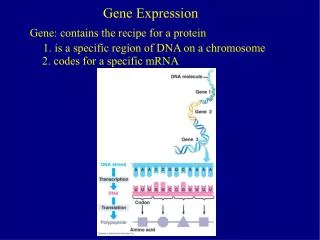
Gene Expression. The human genome contains about 20,325 protein-encoding genes - However, this represents only a small part of the genome Much of the human genome controls protein synthesis - Including the time, speed, and location. Proteins have diverse functions in the body.

Gene Expression
E N D
Presentation Transcript
Gene Expression The human genome contains about 20,325 protein-encoding genes - However, this represents only a small part of the genome Much of the human genome controls protein synthesis - Including the time, speed, and location
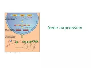
Gene Expression Production of protein from instructions on the DNA Gene expression requires several steps - Transcription = Production of mRNA - Translation = Production of protein using mRNA, tRNA, and rRNA - Folding of the protein into the active 3-D form
Central Dogma The directional flow of genetic information Figure 10.1 Figure 10.1
Transcription RNA is the bridge between DNA and protein RNA is synthesized from one strand of the DNA double helix, which is called the template strand The complementary strand is called the coding strand of DNA Requires the enzyme RNA polymerase
Transcription Figure 10.2 Figure 10.2
Nucleic Acids There are two types of nucleic acids - RNA - DNA Both consist of sequences of N-containing bases joined by sugar-phosphate backbones - However, they differ in several aspects
Nucleic Acids Table 10.2
Figure 10.3 Table 10.2
Types of RNA There are three major types of RNA - messenger RNA or mRNA - ribosomal RNA or rRNA - transfer RNA or tRNA Other classes of RNA control gene expression - Will be discussed in Chapter 11
mRNA Carries information that specifies a particular protein Produced in the nucleus Transported to the ribosome A three nucleotide codon specifies a particular amino acid Most mRNAs are 500-4,500 bases long
rRNA Associate with proteins to make up ribosomes Ribosomes consist of two subunits that join during protein synthesis rRNAs provide structural support - Some are a catalyst (ribozymes) Most rRNAs are from 100-3,000 bases long
rRNA Figure 10.4
tRNA Only 75-80 bases long The 2-D shape is a cloverleaf shape The 3-D shape is an inverted L Has two business ends: - The anticodon forms hydrogen bonds with the mRNA codon - The 3’ end binds the amino acid specified by the mRNA codon
Figure 10.5 Figure 10.6
Transcription Factors In bacteria, operons control gene expression In more complex organisms transcription factors control gene expression and link genome to environment - These contain DNA-binding domains About 2,000 in humans Mutations in transcription factors may cause a wide range of effects
Steps of Transcription Transcription is described in three steps: - Initiation - Elongation - Termination
Steps of Transcription In transcription initiation, a cascade of transcription factors bind to the promoter region of a gene These open a pocket allowing the RNA polymerase to bind just in front of the start of the gene sequence
Steps of Transcription During elongation, RNA polymerase reads the nucleotides on the template strand from 3’ to 5’ and creates an RNA molecule that looks like the coding strand Then termination occurs when sequences in the DNA prompt the RNA polymerase to fall off ending the transcript
Transcription Animation Please note that due to differing operating systems, some animations will not appear until the presentation is viewed in Presentation Mode (Slide Show view). You may see blank slides in the “Normal” or “Slide Sorter” views. All animations will appear after viewing in Presentation Mode and playing each animation. Most animations will require the latest version of the Flash Player, which is available at http://get.adobe.com/flashplayer. Figure 2.3
Simultaneous Transcription of mRNAs Several mRNAs may be transcribed from the same template DNA strand at a time Figure 10.9
RNA Processing In eukaryotes, mRNA transcripts are modified before they leave the nucleus Several steps process pre-mRNA into mature mRNA 1) A methylated cap is added to the 5’ end - Recognition site for protein synthesis 2) A poly-A tail is added to the 3’ end - Stabilizes the mRNA
3) Splicing occurs - Introns (“intervening sequences”) are removed - Exons (“expressed sequences”) are spliced together - Note that introns may outnumber and outsize exons Finally, the mature mRNA is sent out of the nucleus
Figure 10.10 Figure 10.10
Translation The process of reading the mRNA base sequence and creating the amino acid sequence of a protein Occurs on the ribosome Figure 10.11
The Genetic Code The correspondence between the chemical languages of mRNA and proteins In the1960s, researchers used logic and clever experiments with synthetic RNAs to decipher the genetic code More recently, annotations of the human genome has confirmed and extended the earlier work
The Genetic Code It is a triplet code - Three successive mRNA bases form a codon There are 64 codons, including: - One start signal (AUG) - Three stop signals (UAA, UAG, and UGA)
The Genetic Code It is non-overlapping It is degenerate - Two or more codons may specify the same amino acid (synonymous codons) It is universal - Evidence that all life evolved from a common ancestor
Reading Frame A sequence of amino acids encoded from a certain starting point in a DNA/RNA sequence Figure 10.14
Translation Requires mRNA, tRNAs with amino acids, ribosomes, energy molecules (ATP, GTP) and protein factors Divided into three steps: - Initiation - Elongation - Termination
Translation Initiation The leader sequence of the mRNA forms H-bonds with the small ribosomal subunit The start codon (AUG) attracts an initiator tRNA that carries methionine This completes the initiation complex
Translation Elongation The large ribosomal subunit joins A second tRNA binds to the next mRNA codon First peptide bond forms between the two amino acids - Catalyzed by an rRNA ribozyme tRNAs bring in more amino acids, as the ribosome moves down the mRNA - The P site bears the polypeptide chain - The A site holds the newest tRNA
Translation Termination Occurs when a stop codon enters the A site of the ribosome A protein release factor frees the polypeptide The ribosomal subunits separate and are recycled
Translation Animation Please note that due to differing operating systems, some animations will not appear until the presentation is viewed in Presentation Mode (Slide Show view). You may see blank slides in the “Normal” or “Slide Sorter” views. All animations will appear after viewing in Presentation Mode and playing each animation. Most animations will require the latest version of the Flash Player, which is available at http://get.adobe.com/flashplayer. Figure 2.3
Multiple Copies of a Protein Can be Made Simultaneously The closer to the end of the gene, the longer the polypeptide Figure 10.18
Protein Structure Proteins fold into one or more 3-D shapes or conformations There are four levels for protein structure - Primary (1O) structure - Secondary (2O) structure - Tertiary (3O) structure - Quaternary (4O) structure
Protein Folding Protein folding begins as translation proceeds Enzymes and chaperone proteins assist Should a protein misfold, an “unfolded protein response” occurs - Protein synthesis slows or even stops
Protein Misfolding Misfolded proteins are tagged with ubiquitin Then, they are escorted to a proteasome,a tunnel-like multiprotein structure As the protein moves through the tunnel, it is straightened and dismantled Proteasomes also destroy properly-folded proteins that are in excess or no longer needed