Download
1 / 30
310 likes | 707 Views
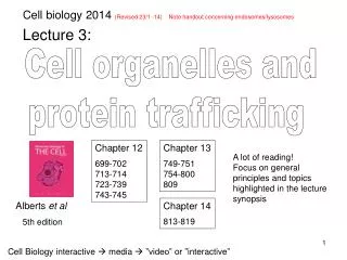
Cell biology 2014 (Revised 23/1 -14) Note handout concerning endosomes/lysosomes. Lecture 3: . Cell organelles and protein trafficking . Chapter 12 699-702 713-714 723-739 743-745. Chapter 13 749-751 754-800 809. A lot of reading!

E N D
Cell biology 2014 (Revised 23/1 -14) Note handout concerning endosomes/lysosomes Lecture 3: Cell organelles and protein trafficking Chapter 12 699-702 713-714 723-739 743-745 Chapter 13 749-751 754-800 809 A lot of reading! Focus on general principles and topics highlighted in the lecture synopsis Albertset al 5th edition Chapter 14 813-819 Cell Biologyinteractive media ”video” or ”interactive”
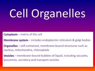
Free cytosolic Ribosome populations Attached to the endoplasmic reticulum Protein trafficking between cell compartments Nucleus ER Golgi Protein N C Ribosomes Cytosol Lysosome Mitochondrion >90 % of all membranes are part of organelles within the cytoplasm Various address tags (without a tag cytosol)
Glucose ATP NADH Pyruvat 3. 1. 2. 2. 1. 3. The cytosol Viscous solution high concentration of proteins (~400 mg/ml) Nucleus Key processes/ components of the cytosol Translation Glycolysis Signal transduction
Reproduce by dividing in two (binary fission) + The mitochondrion - the power plant of the cell • From Greek, mitos, thread, + chondros, granule • The mitochondrion is a double membrane-enclosed organelle • that specialize in ATP regeneration (>100 per cell) 1 mm The invaginations are denoted cristae increased surface area 0.5 mm
Metabolic pathways of the mitochondrion 2. Acetyl CoA Acetyl CoA 1. 2. 1. Pyruvate Fatty acid Krebs cycle Intermediary metabolism NADH Anim. 02.5-citric_acid_cycle.mov (1.5 min) Respiration (electron transport chain) and ATP synthesis Generation of a H+ gradient and utilization of its energy for ATP production (Alberts et al Fig. 14-10) Anim. 14.3-electron_transport & 14.4-ATP_synthase
The origin of the mitochondrion and its genome + Aerobic bacteria ”Founding” eukaryote • Mitochondria have circular DNA and bacteria-like ribosomes 22 tRNA genes 37 genes 2 rRNA genes 13 mRNA encoding genes • Mitochondria are only inherited from the mother • Most of the mitochondrial proteins are encoded in the • nucleus and have to be imported from the cytosol
N Signal sequence Targeting proteins to the mitochondrion Protein translocation across the mitochondrial membranes is mediated by proteins that form a channel spanning both membranes Translocation of mitochondrial proteins through this channel requires proteins to be kept unfolded Folded protein Unfolded protein Chaperone, keeping the protein unfolded in cytosol No passage Successful passage Anim. 12.3-protein_import
Lipid synthesis (Smooth ER) Detoxifications (Smooth ER, eg. P450) storage (Smooth ER) Ca2+ 2. L+i+p+i+d = Lipid Ca2+ 1. 2. 4. 3. 1. 4. 3. Endoplasmic reticulum – ”network within the cell” Protein sorting and modification (Rough ER ) (starting point of the “secretory pathway” of protein synthesis)
Protein targeting to the endoplasmic reticulum Anim. 06.6-translation-I ER associated ribosome SRP receptor Cytosolic ribosome Signal-recognition particle (SRP) Ribosome mRNA tRNA ER lumen Protein translocator ER signal sequence, stretch of hydrophobic a.a. Pause in translation during localization step
Co-translational protein translocation Protein is translocated into the lumen of the ER co-translationally Signal sequence is cleaved by a peptidase after completion of translation/ translocation
Integration of a transmembrane protein into ER C N However, translation continues • Translation complete • the stop-transfer signal sequence integrates into the ER membrane Translocation is initiated but stops at a hydrophobic ~15 aa sequence termed stop-transfer signal
Note the opening of the protein translocater, which allows lateral diffusion within the ER-membrane of both the ER-signal sequence and trans-membrane domains
Synthesis of multi-pass transmembrane proteins Re-start-transfer sequence ER signal sequence (N-terminus)= the initial ”start transfer signal” followed by a signal peptidase recognition site C- Stop-transfer sequence SRP Translocation stop and re-start several times, which results in a multi-pass transmembrane protein Anim. 12.6-protein_translocation.mov
Out of the cell (secretion) Post office Plasma membrane Post ER Lysosome Golgi Secretory pathway Entry into ER is in most cases only the first step to a final destination ER
Post ER Proteins are glycosylated during passage of the secretory pathway “Glycocalyx – a carbohydrate zone on the cell surface” Extracellular Post-translational modification by attachment of oligo-saccharides Cytosol N-linked oligo-saccharides are attached via the amide group of asparagine in ER H N O-linked oligo-saccharides are attached to hydroxyl group of serine or threonine in Golgi Post office O Golgi
Vesicular trafficking post ER Out of the cell Post office Plasma membrane Post ER Lysosome Golgi Secretory pathway Transport from ER to Golgi, within Golgi, and from Golgi to either lysosomes or cell surface is carried out by transport vesicles (liposomes made of phospholipids) Video 13.2-biosy_secret_path
Post ER The architecture of the Golgi apparatus Proteins that keep the Golgi cisterna together Nucleus 3-10 Golgi cisterna (containing different sets of processing enzymes) Trans-face Cis-face Downstream target compartments Transport vesicles
1. 2. 3. Principle of vesicular transport Donor compartment Budding of vesicle from donor compartment The cytoskeleton is used often used as railway tracks Vesicle transport Docking and fusion of a vesicle with its target compartment Target compartment
1. 2. 3. Vesicle formation in donor compartment Vesicle pinching off Vesicle formation Coat protein Bud formation Sorting receptor Constricting protein complex Cargo (i.e., the protein to be transported) Lumen of donor compartment
Clathrin COPI COPII Different coating proteins in vesicular trafficking Endocytosis at the plasma membrane Coat: Lysosome Adaptin Golgi Sorting receptor Cargo ER
1. 2. 3. 1. 2. 3. Vesicle docking and fusion with target compartment Uncoating of vesicle subsequent to ”pinching off” Vesicle tethering with target compartment (specificity Rab’s) Vesicle docking and fusion with target compartment (SNAREs) Lumen of target compartment
Tethering of vesicles to the correct target compartment Rab protein on vesicle docks with Rab effector on target compartment Rab protein Rab effector (tethering protein) Different Rab proteins – different target compartments Compartment X Compartment Y
1. 2. 3. 1. 2. 3. 4. 4. This leads to exclusion of H2O membrane fusion Fusion of a vesicle with its target compartment v-SNARE t-SNARE SNARE proteins on vesicle and target compartment interacts Conformational changes of SNAREs bring the membranes closer together….. …..until they are in physical contact
Protein trafficking in the vesicular pathway Plasma membrane Lysosome Clathrin Golgi COPI COPII Retrieval of ER proteins (KDEL receptor) Retrograde transport Anterograde transport Endoplasmic reticulum
C B A A B C Protein trafficking: post-Golgi Exocytosis Lysosomal pathway Primary lysosome Regulated (e.g. insulin) Constitutive Endosome Secondary lysosome The term lysosome defines a function: lys: digest some: body Lysosomes develop from endosomes by fusion with vesicles carrying lysosomal enzymes Anim. 13.1-clathrin
P The lysosome – the digestive system of the cell • Vesicles (~ 300/cell) filled with ~ 40 acid hydrolases that has • capacity to degrade more or less anything • The lysosome is responsible for degradation of exogenous and • endogenous macromolecules and structures • The inside of the lysosome is acidic pH 7.2 H+ H+ 0.2-0.5 mm H+ pH 5 ATP ADP + H+
The pH regulates the activity of hydrolytic enzymes Lysosome contains many types of hydrolytic enzymes These are only active in an acidic environment + + Degradation of endocytosed material Hydrolases are inactive in ER and Golgi (pH ~7) Hydrolases are active in theacidiclumen of the lysosome Hydrolases: proteases, nucleases, phosphatases etc etc.
1. 2. 3. 1. 2. 3. Uptake of material from the exterior Phagocytosis (“cell eating”) – specific uptake of large (0.5 – 2 mm) particles, primary by immune cells Receptor-mediated endocytosis - specific uptake of molecules Non -specific endocytosis, pinocytosis (“cell drinking”) - anything small in the extracellular fluid istaken up indiscriminately
5. 2. 5. 4. 3. 2. 1. 3. 4. 4. 4. 1. Three routes to the lysosome x Phagocytosis Endocytosis ER Autophagy Phagosome Endosome Autophagosome Primary lysosome Secondary lysosome Anim. 13.3-receptor_endocytosis (Note: vesicle fusion with endosome)
Summary: cellular organelles and trafficking • 3 types of protein transport • A. Gated (nuclear pores) • Across membranes** • (translocation channels) • Vesicle • (budding and fusion) Nucleus (6%) ER (12%) Golgi (3%) Cytosol (54%) ** Ribosomes ** >10-fold more internal membranes than plasma membrane % = volume of a liver cell Lysosome (1%) Mitochondrion (22%) Endosome (1%)