Download
1 / 32
330 likes | 411 Views
Dive into the basic anatomy of the nervous system, covering neuron types, CNS/PNS divisions, functional axon types, and more. Explore the CNS, PNS, glial cells, and functional zones in the neural tube.

E N D
BASIC ANATOMY OF THE NERVOUS SYSTEM
Recommended textbooks: Dubový, Petr. Gross Anatomy and Structure of the Human Nervous System - Part I. Surface Anatomy and Structural Arrangement of the Central Nervous System. 3rd ed. Brno : Masarykova univerzita, 2012. 91 s. ISBN 978-80-210--6125-5. Drake, Richard L. Gray´s anatomy for students. ISBN 9780443069529. Stingl, Grim, Druga: Regional anatomy. Galén 2012, ISBN 978-80-7262-879-7. Dubový, Petr.Instructions for Anatomical Dissection Course. 1. dotisk 2. vyd. Brno: Masarykova univerzita, 2010. 71 s. ISBN 978-80-210-4229-2. Netter, Frank H. Atlas of Human Anatomy, 3rd. ed. 2003. ISBN: 1929007116 Atlas of anatomy:Latin nomenclature. Edited by Anne M. Gilroy - Brian R. MacPherson - Lawrence M. Ross - Michael Schu. New York: Thieme Medical, 2009. xv, 656 p. ISBN 978-1-60406-099-7.
Basic conception afferent information (nerves, axons, tracts) CNS efferent information (nerves, axons, tracts) ascending information (tracts) descending information (tracts)
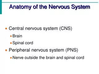
DIVISION OF THE NERVOUS SYSTEM CNS PNS oligodendrocytes astrocytes Schwann cells and their derivatives sensors stimulus impulse effectors Striated skeletal muscles Non-striated muscles, myocardium, glands
DIVISION OF THE PNS Cranial nerves I.- XII. • run through the skull base Spinal nerves – 31 pairs • run through foramina intervertebralia
Glialcells of the CNS: astrocytes, oligodendrocytes, microglial, ependymal cells Glial cells of thePNS: myelinating andnon-nemyelinating Schwann cells, satellite glialcells, terminal glial cells
unmyelinated axons (< 1m) myelinated axons
FUNCTIONAL TYPES OF AXONS IN PNS somatosensory touch, proprioception, pain viscerosensory mechanoception, pain Afferent relay impulses for taste, hearing and balance sensory somatomotor striated muscles striated muscles branchiomotor Efferent non-striated muscles visceromotor sympathetic myocardium parasympathetic glands
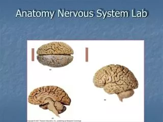
DIVISION OF THE CNS Brain (Encephalon) Spinal cord (Medulla spinalis) Brainstem (Truncus encephali) Medulla oblongata Pons Mesencephalon Cerebellum Diencephalon Telencephalon
Primary subdivisions: prosencephalon, mesencephalon, rhombencephalon myelencephalon metencephalon mesencephalon diencephalon medulla spinalis telencephalon Secondary subdivision: telencephalon, diencephalon, mesencephalon, metencephalon, myelencephalon
Conus medullaris Filum terminale Cauda equina
Spinal segment Fila radicularia
Intumescentia cervicalis C3 – T2 Intumescentia lumbalis T9 – T12
Segment C5 Segment C1 Segment C5 Segment C8 Segment C8 Segment Th2 Segment Th2 Segment Th10 Segment L4 Segment L1 Segment S4 Segment L4 Segment S4
SUBSTANTIA GRISEA – cornu anterius (columna anterior), cornu posterius (columna posterior), cornu laterale (columna lateralis), substantia intermedia, canalis centralis
SUBSTANTIA ALBA – funiculus anterior, lateralis, posterior fissura mediana ant., sulcus medianus post., septum medianum posterius, sulcus anterolateralis, posterolateralis
FUNCTIONAL ZONES IN THE NEURAL TUBE S SS VS cc VM SM BM
Cornu posterius FR Substantia intermedia CC Cornu anterius
Pseudounipol. neurons of the DRG Radix dorsalis Tr. dorsolateralis Lissaueri Ncl. posteromarginalis + Subst. gelatinosa Rolandi Fasc. gracilis Tr. spinobulbaris Fasc. cuneatus Ncl. proprius Ncl. thoracicus (Stilling-Clark.) Tr. spinocerebellaris post. Ncl. intermediomedialis Tr. spinocerebellaris ant. Tr. spino-thalamicus, -reticularis, -tectalis Tr. spino-olivaris Tr. olivo-spinalis
Ncl. intermediolateralis Ncl.motorius
Tr. corticospinalis lat. Tr. rubrospinalis Tr. reticulospinalis Tr. olivospinalis Tr. tectospinalis Tr. corticospinalis ant. Tr. vestibulospinalis