Download
1 / 57
771 likes | 1.59k Views
The Anatomy of the Nervous System. Why study Neuroanatomy? . Major divisions of the nervous system. Major divisions of the nervous system. Brain. Central nervous system. Spinal cord. Afferent nerves. Sensory CNS info. Nervous System. Somatic nervous system.

E N D
The Anatomy of the Nervous System Why study Neuroanatomy?
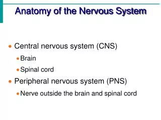
Major divisions of the nervous system Brain Central nervous system Spinal cord Afferent nerves Sensory CNS info Nervous System Somatic nervous system CNS Skeletal muscles Efferent nerves Peripheral nervous system Afferent nerves Autonomic nervous system Parasympathetic nervous system Efferent nerves Sympathetic nervous system
The spinal cord Cross section through the spinal cord Ventral horn
Cranial Nerves • Olfactory (smell) • Optic (vision) • Oculomotor (eye movement) • Trochlear (eye movement) • Trigeminal (facial sensation and chewing) • Abducens (eye movement) • Facial (taste and facial expression) • Auditory (hearing and balance) • Glossopharyngeal (taste, salivation swallowing) • Vagus (abdominal organs, throat muscles) • Spinal accessory (neck, shoulders, head) • Hypoglossal (tongue)
http://www.wnyc.org/shows/radiolab/episodes/2006/05/05 The body-brain connection • Radio Lab 2006 • Where Am I? • Mind and body are in constant communication (neuroscientists call this the brain-body loop), but the loop can get out-of-sync-- even broken. This hour: stories of people whose brains and bodies have lost each other. We begin with a century-old mystery: why do many amputees still feel their missing limbs? We speak with a neuroscientist who solved the problem with a magician's trick: an optical illusion. We continue with the story of a butcher who suddenly lost his entire sense of touch, and how, after many years, he managed to grow a new sense. And we hear from pilots who lose consciousness and suffer out-of-body experiences while flying fighter jets.
The Anatomy of the Nervous System Neuroanatomy cont.
Anatomical Directions Rostral Caudal Sagittal plane (midsagittal section) Horizontal plane Frontal plane Cross section (coronal section)
Five Major divisions of the Brain ] • Telencephalon • Diencephalon • Mesencephalon • Metencephalon • Myelencephalon Forebrain ] Midbrain Brainstem ] Hindbrain
The Hindbrain • Myelencephalon (medulla) • Tracts • Small nuclei • Reticular formation (RT) • Metencephalon • Cerebellum (little brain) • Pons (bridge) • Neural tracts
Mesencephalon - The Midbrain Tectum • Superior collicui (visual relay) #2 • Inferior collicui (auditory relay) #3
Mesencephalon - The Midbrain Tegmentum • RF & tracts of passage • Periaqueductal gray • Substantia nigra • Red nucleus
Mesencephalon - The Midbrain • Periaqueductal gray - mediates the analgesic effects of opiate drugs. • Substantia nigra (black substance) – neurons project to striatum; degenerate in PD. • Red nucleus – motor pathways from cortex and cerebellum.
Diencephalon • Thalamus – large two-lobed structure; is the top of the brain stem. Contains many different nuclei, most of which project to the cortex • Sensory relay nuclei • Massa intermedia • White lamina
Diencephalon • Hypothalamus – Below the anterior thalamus. Regulates several motivated behaviors. • Pituitary gland • Optic chiasm • Mammillary bodies • Other nuclei • LH (lateral H.) • VMH (ventromedial H.) LH VMH
Telencephalon • Cerebral cortex • Major fissures • Major gyri • Four lobes • Limbic system • Basal ganglia • Cerebral commissures
Telencephalon • Cerebral cortex • Major fissures • Lateral fissure • Central fissure • Longitudinal fissure
Telencephalon Cerebral cortex Rat brain (Lissencephalic)
Telencephalon Cerebral cortex of human, chimpanzee and rat
Telencephalon • Cerebral cortex • Major fissures • Major gyri • Four lobe • Frontal • Parietal • Temporal • Occipital
Telencephalon • 90% of the cortex in Humans is neocortex, which has 6 distinct cell layers. As the name implies, Neo-cortex is a more recent development of brain evolution.
Telencephalon • Hippocampus – it is cortex, but not neo-cortex (it only has 3 layers). It is sometimes called Archicortex. • Can you see the Sea Horse?
Telencephalon – Subcortical parts Basal Ganglia Limbic System
Telencephalon – Limbic system • Hippocampus • Amygdala • Fornix • Septum • Cingulate cortex • Mammilary bodies
Telencephalon – Basal Ganglia • Caudate nucleus • Putamen • Globus pallidus • Amygdala • Substantia nigra • Subthalamic n. • thalamus • cortex
Telencephalon – Basal Ganglia • Caudate nucleus • Putamen • Globus pallidus • Amygdala • Substantia nigra • Subthalamic n. • thalamus • cortex
Telencephalon – Basal Ganglia • Caudate nucleus • Putamen • Globus pallidus • Amygdala • Substantia nigra • Subthalamic n. • thalamus • cortex
Name the brain region 1. Type of section? 7. • Mid-saggital • Cingulate • Corpus callosum • Pons • Temporal • Cerebellum • Parietal • Occipital • Frontal • Thalamus • Tegmentum • Tectum • Hypothalamus • Mammillary • bodies 2. 3. 9. 8. 10. 12. 13. 14. 11. 6. 5. 4.
Meninges • Dura mater (tough mother)outer membrane. • Arachnoid membrane (web-like) a thin membrane. • Subarachnoid space – contains large blood vessels and CSF. • Pia mater (pious or gentle mother) adheres to the surface of the CNS.
Ventricles & CSF • Cerebral Ventricles Four large internal chambers of the brian. • Lateral ventricles, 3rd ventricle, & 4th ventricle. • Central canal – a small canal that runs the length of the spinal cord. • Choroid plexuses are a network of capillaries that protrude into the ventricles and produce cerebrospinal fluid (CSF).
Lateral ventricles • Third ventricle • Cerebral aqueduct • Fourth Ventricle • Arachnoid villi • Choroid plexus • Choroid plexus • Subarachnoid space
Protecting the Brain Physical protection • Skull & Vertebrae • Meninges • Cerebrospinal fluid (CSF) Chemical protection • Blood-brain barrier (BBB)
Blood-brain barrier • Results from the special structure of cerebral blood vessels. • Cells in the walls of cerebral blood vessels are tightly packed. • This provides a barrier for the passage of some large-molecules and proteins into the brain. • Not all large molecules are impeded (e.g., glucose). • Sex hormones readily pass through to certain brain areas where the BBB is weak.
Two basic cells of the nervous system • Neurons –cells specialized for the reception, conduction and transmission of electrochemical signals. • Glial cells – classic view - support cells that a) provide nutrients b) clear waste c) provide a physical matrix (glia means “glue”) But recent evidence suggests that they do even more…
Two basic cells of the nervous system Glial cells also – • Participate in neurotransmission by sending signals to neurons and receiving signals from them. • Control the establishment and maintenance of synapses • Form circuits and may contribute to synaptic plasticity.
External Anatomy of the Neuron • Cell body (soma) • Cell membrane • Dendrites • Axon • Axon hillock • Myelin • Nodes of Ranvier • Terminal boutons • Synapses