Download
1 / 25
250 likes | 580 Views
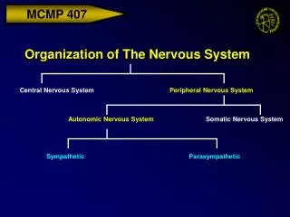
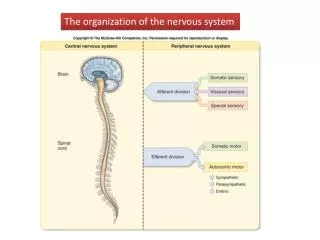
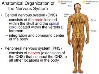
The basic organization of the nervous system. The divisions of the nervous system. The nervous system is composed of • central nervous system • the brain • spinal cord • peripheral nervous system • somatic nervous system - afferent nerves - efferent nerves

E N D
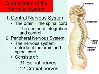
The divisions of the nervous system The nervous system is composed of • central nervous system • the brain • spinal cord • peripheral nervous system • somatic nervous system - afferent nerves - efferent nerves • autonomic nervous system - sympathicus - parasympathicus
Dorsal and ventral root The mixed nerve splits into the dorsal and the ventral root. The dorsal root contains the sensory axons. The ventral root contains the motor axons.
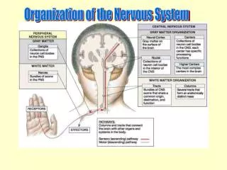
The spinal cord • White matter forms the external part of the cord. It is made up of myelinated axons. These carry signals up and down the axis of the spinal cord, and make it white. • Gray matter forms the butterfly shaped inner core of the cord. It is made up of neuron cell bodies, dendrites and synapses. Here neurotransmission takes place.
White and gray matter The spinal cord contains inputs and outputs (the spinal nerves) it has ascending and descending axon tracts passing through it (some of which form synaptic terminals) and it has collections of neurons (or nerve cell bodies) that process information and perform functions
Some properties of the spinal cord • Signals enter and leave the spinal cord via spinal nerves. Of these pair exists for each spinal vertebra. They are mixed nerves carrying sensory information to the spinal cord and motor information from the spinal cord to the muscles. • The lower and upper limbs require many more sensory and motor axons than the chest and abdomen. For this reason the spinal cord is larger in the lumbar and cervical regions than in the thoracic region. • The spinal cord ends at about L1 and the lumbar nerves descend within a CSF filled sack down to their point of exit. This fluid space caudal to the end of the spinal cord is called the lumbar cistern and is the point that CSF can be sampled using lumbar puncture.
The central nervous system The brain is composed of • the brainstem (rombencephalon & mesencephalon) • the cerebellum • the diencephalon • the telencephalon (cerebrum)
The brainstem • consists of the medulla oblongata (caudal) the pons (middle) and the midbrain (rostral). • connects to the spinal cord at its caudal end and to the diencephalon at its rostral end. • like the spinal cord, the brainstem spinal cord contains inputs and outputs, the cranial nerves. It has ascending and descending axon tracts passing through it. Some of these form synaptic terminals. Furthermore, it has collections of neurons that process information and perform functions.
Medulla oblongata • consists of the medulla oblongata (caudal) the pons (middle) and the midbrain (rostral). • connects to the spinal cord at its caudal end and to the diencephalon at its rostral end. • like the spinal cord, the brainstem spinal cord contains inputs and outputs, the cranial nerves. It has ascending and descending axon tracts passing through it. Some of these form synaptic terminals. Furthermore, it has collections of neurons that process information and perform functions.
The cranial nerves • spinal nerves carry sensory and motor information from the level of the sacrum and coccyc (sacral and coccygeal nerves) to the level of the neck (upper cervical nerves) • above this level, in the region of the head and face, 12 nerves enter the cranium rather than the spinal cord and are termed cranial nerves
Pyramidsthe corticospinal tract fibers within the medulla form a distinct feature called the pyramids, they decussate, which means that the axons within this fiber bundle cross from the left side of the brain to the right side of the brain. The cortex on the right side of the brain send and receives information from the left side of the body. I do not know why this is the case but it means that ascending sensory information and descending motor information has to deccusate (cross the midline) between the cerebral cortex and the spinal cord. Dorsal column nuclei – sensory axons that do not synapse in the spinal cord dorsal horn ascend the spinal cord in the dorsal column nuclei and synapse in dorsal column nuclei in the medulla Cranial nerve nuclei – these are nuclei (collections of cell bodies) within the medulla that receive sensory information from cranial nerves (like the dorsal horn of the spinal cord) or are collections of motor neurons that send out information to muscles of the head and face (like the ventral horn of the spinal cord).
The pons • is connected to the medulla caudally and to the midbrain rostrally. • Pons means “bridge” in Latin and it referes to the fact the ventral surface of the pons looks like a bridge because of the massive numbers of fibers (myelinated axons) that decussate (cross the midline) in this part of the brainstem. These crossing fibers are axons which project from the pons to the cerebellum. • The pons contains inputs and outputs (some cranial nerves) it has ascending and descending axon tracts passing through it (some of which form synaptic terminals) and it has collections of neurons (or nerve cell bodies) that process information and perform functions.
The cerebellum The cerebellum ("little brain") has convolutions similar to those of cerebral cortex, only the folds are much smaller. Like the cerebrum, the cerebellum has an outer cortex, an inner white matter, and deep nuclei below the white matter.
If we enlarge a single fold of cerebellum, or a folium, we can begin to see the organization of cell types. The outermost layer of the cortex is called the molecular layer, and is nearly cell-free. Instead it is occupied mostly by axons and dendrites. The layer below that is a monolayer of large cells called Purkinje cells, central players in the circuitry of the cerebellum. Below the Purkinje cells is a dense layer of tiny neurons called granule cells. Finally, in the center of each folium is the white matter, all of the axons traveling into and out of the folia. These cell types are hooked together in stereotypical ways throughout the cerebellum.
The mesencephalon (midbrain) The upper part of the brainstem is called the midbrain It also contains inputs and outputs (some cranial nerves) it has ascending and descending axon tracts passing through it (some of which form synaptic terminals) and it has collections of neurons (or nerve cell bodies) that process information and perform functions. The most notable anatomical features of the midbrain are: Cerebral peduncles – contain the descending corticospinal fibers Medial lemniscus – contain the ascending sensory fibers Superior (and inferior) colliculus – contain visual orientation centers Substantia nigra – very important nuclei for motor control
The diencephalon The rostral part of the brainstem merges with the diencephalon. thalamus – the part of the brain that recieves synapses from all ascending sensory innervation and then sends a projection to the appropriate part of the cerebral cortex. hypothalamus - the command center for homeostasis within the brain. controlling blood pressure, body temperature, blood glucose and all of the other ‘housekeeping’ functions of the body. Only one cranial nerve enters at the level of the diencephalon and that is the optic nerve carrying visual sensory information. Ascending and descending information within the diencephalon travels in axon tracts within the internal capsule.
The telencephalon Cerebrum The cerebrum is the most rostral part of the central nervous system. It is compose of the Cerebral cortex Basal ganglia
The cerebral cortex Cortex - the outer coating of what we think of as the brain. It is in many animals convoluted into ridges (gyri) and folds (sulci).
The cerebral lobes The cerebral cortex is organized into territories with different functions called lobes. • The frontal lobe sends motor commands to the brainstem and spinal cord. • The parietal lobe receives somato-sensory information (pain and temperature, touch and pressure) • The occipital lobe is the site of receipt of visual information. • The temporal lobe is site of receipt of auditory information.
The basal ganglia Generally a ganglion is a collection of cell bodies outside the central nervous system. Not here:he basal ganglia are a collection of nuclei deep to the white matter of cerebral cortex. • The name includes: caudate, putamen, nucleus accumbens, globus pallidus, substantia nigra, subthalamic nucleus. • In principlethe claustrum and the amygdala are part of this collection. However they do not really deal with movement, nor are they interconnected with the rest of the basal ganglia, so they have been dropped from this section. • Obsolete, but are still encountered: the striatum (caudate + putamen + nucleus accumbens), the corpus striatum (striatum + globus pallidus), or the lenticular nucleus (putamen + globus pallidus).
A first look at systems interaction The basal ganglia and cerebellum are large collections of nuclei that modify movement on a minute-to-minute basis. Motor cortex sends information to both, and both structures send information right back to cortex via the thalamus. (Remember, to get to cortex you must go through thalamus.) The output of the cerebellum is excitatory, while the basal ganglia are inhibitory. The balance between these two systems allows for smooth, coordinated movement, and a disturbance in either system will show up as movement disorders.