Download
1 / 32
320 likes | 366 Views
Detailed classification of hypersensitivity reactions (Types I-IV) and mechanisms involved. Covers symptoms, mechanisms, and treatment options for Type I, II, and III reactions.

E N D
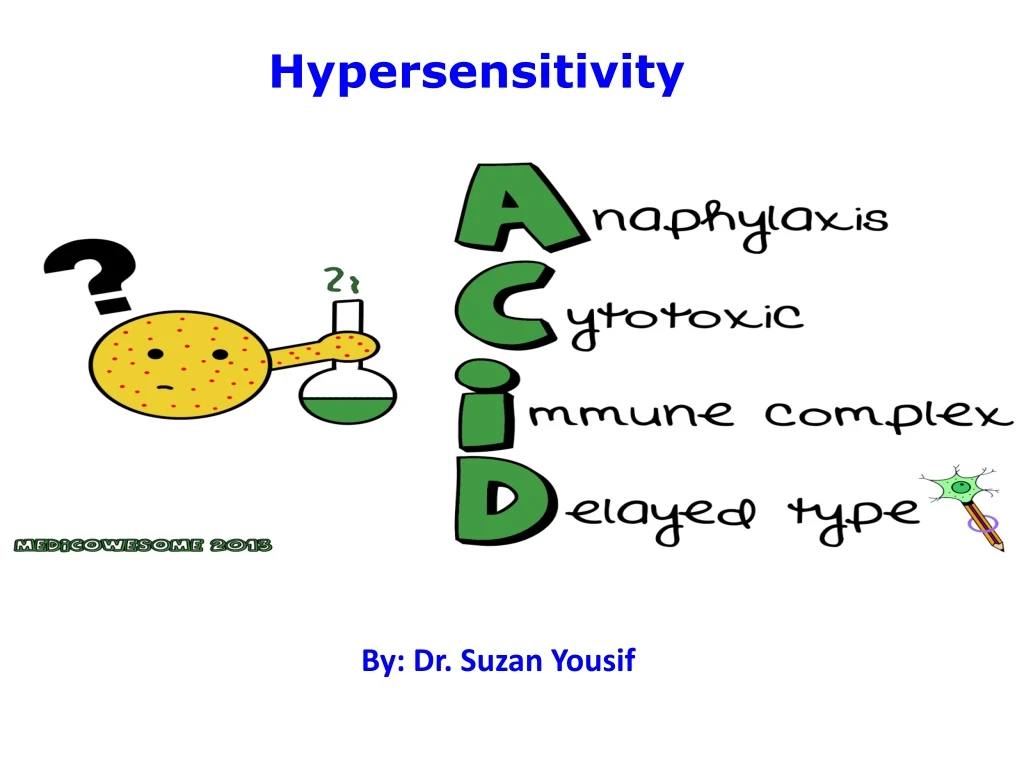
Hypersensitivity By: Dr. Suzan Yousif
Classification • Coombs and Gell classification 1-Type I - anaphylactic( atopic, or immediate) 2-Type II - cytotoxic (antibody-dependent) 3-Type III - immune complex 4-Type IV - delayed orcell-mediated
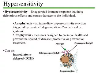
TYPE I HYPERSENSITIVITY Type I hypersensitivity is also known as immediate or anaphylactic hypersensitivity. The reaction may involve skin (urticaria and eczema), eyes (conjunctivitis), nasopharynx (rhinitis), bronchopulmonary tissues (asthma) and gastrointestinal tract (gastroenteritis)
The reaction may cause a range of symptoms from minor inconvenience to death. • The reaction usually takes 15 - 30 minutes from the time of exposure to the antigen. • sometimes it may have a delayed onset (10 - 12 hours).
Immediate hypersensitivity is mediated by IgE. • The primary cellular component in this hypersensitivity is the mast cell or basophil. • The reaction is amplified and/or modified by platelets, neutrophils and eosinophils. • A biopsy of the reaction site demonstrates mainly mast cells and eosinophils.
Mechanism: • The mechanism of reaction involves preferential production of IgE, in response to certain antigens (allergens). • IgE has very high affinity for its receptor on mast cells and basophils. • A subsequent exposure to the same allergen cross links the cell-bound IgE and triggers the release of various pharmacologically active substances like histamine, heparin tryptase, chymase, and monocyte chemotactic factors.
Contents of the Mast Cell Granules Active agent Activity Histamine Increases vascular permeability; elevates level of cyclic AMP Heparin Anticoagulation Serotonin Increases vascular permeability Chymase Proteolysis Hyaluronidase Increases vasuclar permeability Eos. Chem. Factor Chemoattraction of eosinophils Neut. Chem. Factor Chemoattraction of neutrophils Platelet Agg. Factor Aggregates platelets
Localized anaphylaxisTarget organ responds to direct contact with allergen. • Digestive tract contact results in vomiting, cramping, diarrhea. • Skin sensitivity usually inflamed area resulting in itching. • Airway sensitivity results in sneezing and rhinitis OR wheezing and asthma.
Systemic anaphylaxis • Systemic vasodilation and smooth muscle contraction leading to severe bronchiole constriction, edema, and shock. • Similar to systemic inflammation.
Treatment for Type I • Pharmacotherapy Drugs. • Non-steroidal anti-inflammatories • Antihistamines block histamine receptors. • Steroids • Theophylline OR epinephrine -prolongs or increases cAMP levels in mast cells which inhibits degranulation.
Treatment for Type I • Immunotherapy • Desensitization (regular administration of gradually increasing doses of allergen extracts over a period of years) • Repeated injections of allergen to reduce the IgE on Mast cells and produce IgG.
TYPE II HYPERSENSITIVITYcytotoxic • Type II hypersensitivity is also known as antibody-dependent hypersensitivity and may affect a variety of organs and tissues. • In type II hypersensitivity, the antibodies produced by the immune response bind to antigens on the patient's own cell surfaces. • The antigens recognized in this way may either be intrinsic ("self" antigen, innately part of the patient's cells) or extrinsic (absorbed onto the cells during exposure to some foreign antigen, possibly as part of infection with a pathogen
IgG and IgM antibodies bind to these antigens to form complexes that activate the classical pathway of complement activation for eliminating cells presenting foreign antigens • As a result mediators of acute inflammation are generated at the site and membrane attack complexes cause cell lysis and death. The reaction takes hours to a day.
Examples • Autoimmune haemolytic anaemia • Pernicious anemia • Immune thrombocytopenia • Transfusion reactions • Hashimoto's thyroiditis • Graves' disease • Myasthenia gravis • Hemolytic disease of the newborn
Hemolytic disease of newborn Rh factor incompatibility • IgG abs to Rh an innocuous RBC antigen • Rh+ baby born to Rh-mother first time fine. 2nd time can have abs to Rh from 1st pregnancy. • Ab crosses placenta and baby kills its own rbcs. • Treat mother with ab to Rh antigen right after birth and mother never makes its own immune response.
TYPE III HYPERSENSITIVITY immune complexes • Large amount of antigen and antibodies form complexes in blood. • If not eliminated can deposit in Kidneys, Joints, Lung, Skinand trigger inflammation. • PMNs and macrophages bind to immune complexes via FcR and phagocytize the complexes. • If unable to phagocytize the immune complexes can cause inflammation via C’ activation.
Serum sickness • Is a disease caused by the injection of large doses of a protein antigen into the blood and characterized by the deposition of antigen-antibody complexes in blood vessel walls, especially in the kidneys and joints.
Systemic Lupus Erythmatosus • The disease is characterized by the presence of autoantibodies , which form immune complexes with autoantigens and are deposited within the kidney glomeruli • The resulting type III hypersensitivity is responsible for the glomerulonephritis (Inflammation of blood capillary vessels in the glomeruli)
Delayed type hypersensitivity TYPE IV HYPERSENSITIVITY • DTH response is from: • Th1 cells release cytokines to activate macrophages causing inflammation and tissue damage. • Continued macrophage activation can cause chronic inflammation resulting in tissue lesions, scarring, and granuloma formation. • Delayed is relative because DTH response arise 24-72 hours after exposure rather than within minutes. • Unlike the other types, it is not antibody mediated but rather is a type of cell-mediated response.
Stages of Type IV DTH Sensitization stage • Memory Th1 cells against DTH antigens are generated by dendritic cells during the sensitization stage. • These Th1 cells can activate macrophages and trigger inflammatory response.
Stages of Type IV DTHEffector stage • Secondary contact yields what we call DTH. Th1 memory cells are activated and produce cytokines.which cause tissue destruction, inflammation. • Inflamed area becomes red and fluid filled can form lesion. • Continued exposure to antigen can cause chronic inflammation and result in granuloma formation.
Delayed type hypersensitivity (DTH) DTH can be the result of Chronic infection or Exposure to some antigens.
Type IV DTH Contact dermatitis • The response to poison oak is a classic Type IV. • Small molecules act as haptens and complex with skin proteins to be taken up by APCs and presented to Th1 cells to get sensitization. • During secondary exposureTh1 memory cells become activated to cause DTH.