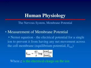
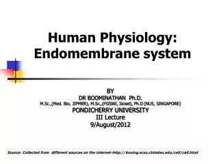
Human Physiology: Endomembrane system
Human Physiology: Endomembrane system. BY DR BOOMINATHAN Ph.D. M.Sc.,(Med. Bio, JIPMER), M.Sc.,(FGSWI, Israel), Ph.D (NUS, SINGAPORE) PONDICHERRY UNIVERSITY III Lecture 9/August/2012.

Human Physiology: Endomembrane system
E N D
Presentation Transcript
Human Physiology:Endomembrane system BY DR BOOMINATHAN Ph.D. M.Sc.,(Med. Bio, JIPMER), M.Sc.,(FGSWI, Israel), Ph.D (NUS, SINGAPORE) PONDICHERRY UNIVERSITY III Lecture 9/August/2012 Source: Collected from different sources on the internet-http://koning.ecsu.ctstateu.edu/cell/cell.html
Learning objectives 1.The structure and function of endoplasmic reticulum(ER); 2.The structure and function of Golgi complex; 3.The structure and function of lysosomes .
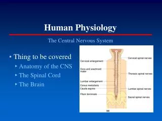
Introduction The Compartmentalization in Eukaryotic Cells • Membranes divide the cytoplasm of eukaryotic cells into distinct compartments. Three categories in eukaryotic cells: 1.the endomembrane system: endoplasmic reticu- lum, Golgi complex, Lysosomes. 2.the cytosol. 3.mitochondria, peroxisomes and the nucleus. Membrane-bound structures (organelles) are found in all eukaryotic cells.
Introduce • Endomembrane System : The structural and functional relationship organelles include endoplasmic reticulum ,Golgi complex, lysosome, secretory vesicles. • Membrane-bound structures (organelles) are found in all eukaryotic cells.
Endomembrane System endoplasmic reticulum(ER) Golgi complex lysosome secretory vesicles
Section IEndoplasmic Reticulum 1.1 The Structure of ER The endoplasmic reticulum is a network of flattened sacs and branching tubules that extends throughout the cytoplasm in plant and animal cells. These sacs and tubules are all interconnected by a single continuous membrane so that the organelle has only one large, highly convoluted and complexly arranged lumen (internal space).
1.2 The types of ER The ER comes in two forms. Rough endoplasmic reticulum(RER) Smooth endoplasmic reticulum(SER)
1.2 The types of ER 1.2.1 The rough ER The ER are covered with ribosome .Their rough appearance under electron microscopy led to their being called rough ER . RER has ribosomes on the cytosolic side of continuous, flattened sacs.
1.2 The types of ER 1.2.1 The rough ER The outer membrane of the nucleus is always studded with ribosomes and is continuous with rough ER membrane . The lumen of RER is connected to nuclear envelope .
1.2 The types of ER 1.2.2 The smooth ER The parts are free of ribosomes and are called smooth ER (SER). SER is an interconnecting network of tubular membrane elements.
1.2 The types of ER • Rough and smooth ER differ not only in structure, but also in function.
1.3 The functions of the RER 1.3.1 Proteins synthesized on ribosomes of RER : * The ribosomes assemble amino acids into protein units, which are transported into the lumen of rough endoplasmic reticulum for further processing. * These proteins may be either transmembrane proteins, which become embedded in the membrane of the endoplasmic reticulum, or water-soluble proteins, which are able to pass completely through the membrane into the lumen.
1.3 The functions of the RER Those proteins that reach the inside of the endoplasmic reticulum are folded into the correct three-dimensional conformation. Chemicals, such as carbohydrates or sugars, are added, then the ER either transports the completed proteins to areas of the cell where they are needed, or they are sent to the Golgi apparatus for further processing and modification.
1.3 The functions of the RER 1.3.1 Proteins synthesized on ribosomes of rER include: • Secretory proteins; • integral membrane proteins; • soluble proteins of organelles.
1.3 The functions of the RER ? How do the ribosomes (a membrane-bound ribosome ) attach to the outer surface of rER ,and then the newly synthesized protein pass completely through the membrane into the lumen of rER?
1.3 The functions of the RER The Signal Hypothesis would explain completely the mechanism that the ribosomes attach to the outer surface of rER ,and then the newly protein pass completely through the membrane into the lumen of rER?
1.3 The functions of the RER The basic structure of the process is a Signal Reco-gnition Particle(SRP),that lead the ribosomes attach to the outer surface of rER ,and then lead the newly protein pass completely through the membrane into the lumen of rER. SRP
1.3 The functions of the RER • Signal-recognition particle(SRP)1.synthesis:Six different polypeptides complexed with a 300-nucleotide molecule of RNA.
1.3 The functions of the RER 2. SRP have three main active sites:* One that recognizes and binds to ER signal sequence;* One that interacts with the ribosome to block further translation;* One that binds to the ER membrane (docking protein:receptor protein) The site to recognize and bind to ER signal sequence The site to block further translation The site to recognize receptor protein
1.3 The functions of the RER • ER signal sequence:1. synthesis :Typically 15-30 amino acids.2.consist of three domains: a positively charged N-terminal region;a central hydrophobic region;a polar region adjoining the site where cleavage from the mature protein will take place. A signal sequence on nascent seretory proteins targets them to the ER and is then cleaved off.SRP receptor(a binding protein or docking protein:receptor protein)
Signal peptides and Signal patches Figure6-8Two ways that a sorting signal can be built into a protein.(A) The signal resides in a single discrete stretch of amino acid sequence, called a signal peptide, that is exposed in the folded protein. Signal peptides often occur at the end of the polypeptide chain, but they can also be located elsewhere. (B) A signal patch can be formed by the juxtaposition of amino acids from regions that are physically separated before the protein folds; alternatively, separate patches on the surface of the folded protein that are spaced a fixed distance apart could form the signal.
1.3 The functions of the RER The basic structure of the mechanism of Signal Hypothesis :1. Signal Recognition Particle(SRP)2. ER signal sequence 3.SRP receptor 1 3 SRP
The mechanism of the Signal Hypothesis As thesignal sequenceemerge from the ribosome, they are recognized and bound by asignal recognition particle(SRP), This binding inhibits translation and target the complex to the RER by binding to theSRP receptoron the ER membrane. SRP is then released and the ribosome binds to aprotein translocation complexin the ER membrane; the signal sequence is inserted into a membrane channel, translation is resumed and the unfolded growing polypeptide chain is translocated across the membrane into the ER. As translocation proceeds, the signal sequence is cleaved bysignal peptidaseand the polypeptide is released into the ER lumen.
Signal Hypothesis (SRP) mRNA A P A ribosome signal sequence tRNA Channel protein核糖体结合蛋白 SRP receptor cytoplasme The lumen of RER
This process shows the signal receptor particle that associates with the large and small subunit of the ribosome that allows binding to the receptor on the rough endoplasmic reticulum.After the protein is synthesized, the ribosome dissociates into large and small subunits and the SRP also looses its attachment to the receptor.
1.3.2 Modification/processing of newly synthesized proteins: glycosylation in theRER Glycosylation of newly synthesized proteins • N-linked: oligosaccharide chain islinked to the amide nitrogen of asparagine (Asn) (in ER) • O-linked: oligosaccharide chain islinked to the hydroxyl group of serine or threonine (in Golgi)
1.3.2N-linked glycosylation in the RER Lipid-linked oligosaccharide chain is added to the dolichol by glycosyltransferase,then be transferred to linked to the N terminus ofasparagines within polypeptide by oligosaccharine protein transferase.
1.3.2 Modification/processing of newly synthesized proteins: the folding of proteins The lumen of rER contains: These chaperones and enzymes recognize and bind to unfolded or misfolded proteins and give them correct conformation; Quality control: ensuring that misfolded proteins do not leave ER.
1.3.2 Modification/processing of newly synthesized proteins : the formed of disulfide bonds within polypeptide • The lumen of rER contains: • Protein disulfide isomerase ( PDI ) transfer incorrect disulfide bonds to correct disulfide bonds within polypeptide.
1.3.3 The transport of the proteins • The transport of the • proteins contains: • 1.The formation of transport • Vesicle (secretory proteins); • 2.The transport of • integral membrane proteins;3.The transport ofsoluble proteins of organelles.
1.3 The functions of the RER 1.Proteins synthesized on ribosomes of RER. 2.Modification and processing of newly synthesized proteins ; A.glycosylation in the RER.B. the folding of proteins.C. the formation of disulfide bonds within polypeptide.3. The transport of the proteins .
1.4 Functions of the SER The side of cytoplasm 1.It takes part in the synthesis of various lipids: phospholipids (building membranes ) fatty acids steroids (e.g.,hormones). The lumen of SER flipase
1.4 Functions of the SER • Transport by phospholipid exchange proteins (PEP):SER→other organelles. phospholipid exchange proteins (PEP)
1.4 Functions of the SER 2.Detoxification of organic compounds in liver cells. It contains enzymes needed to detoxify drugs. 3.Metabolism of heparin. 4.The SER serve as a storage place for calcium.
The SER serve as a storage place for calcium In the case of smooth endoplasmic reticulum in muscle cells, the tubules serve as a store of calcium which is released as one step in the contraction process of muscle. Calcium pumps serve to move the calcium.
1.4 Functions of the SER 1.Synthesis of lipid; 2.Detoxification of organic compounds in liver cells; 3.Metabolism of heparin; 4.The SER serve as a storage place for calcium.
What are the functional differences between the RER and the SER? Functions of the RER 1.Proteins synthesized on ribosomes of RER. 2.Modification and processing of newly synthesized proteins ; A.glycosylation in the RER.B. the folding of proteins.C. the formation of disulfide bonds within polypeptide.3. The transport of the proteins . Functions of the SER 1.Synthesis of lipid; 2.Detoxification of organic compounds in liver cells; 3.Metabolism of heparin; 4.The SER serve as a storage place for calcium.
Summary • ER is a network of folded membranes that extend through the cytoplasm to the nuclear membrane. • There are two kinds of ER, rough and smooth. • The functions of RER include the synthesis of protein, modification/processing and quality control of newly synthesized proteins. • The SER has functions in several metabolic processes. • It takes part in the synthesis of various lipids , fatty acids and steroids, and also plays an important role in carbohydrate metabolism, detoxification of the cell and calcium storage.
Self-Quiz :Choosed-question 1.Where does protein synthesis take place in eucaryotic cells ? A.granum B.nucleus C.endoplasmic reticulum D.golgi apparatus
Self-Quiz 2 .The ribosomes of prokaryotic cells are found A.in the golgi apparatus B.free-floating C.in the nucleus D.in the endoplasmic reticulum
Self-Quiz 3.The smooth endoplasmic reticulum is the area in a cell where ___ are synthesized. A.polysaccharides; B.proteins; C.lipids; D.DNA
SectionⅡ The Golgi complex The Golgi apparatus is a polarized struc-ture consisting of an oriented stack of disc-shaped cisternae sur-rounded by a swarm of small vesicles.
2.1 The structure of Golgi complex • The Golgi complex consists of a stack of flattened、 vesiclesandtubules. • The Golgi apparatus has two distinct faces: • a cis ,or forming face • a trans,or maturing face
2.2 The polarity of Golgi complex The cis face is closely associated with a transitional RER. In secretory cells ,the trans face is the closest to the plasma membrane.The large secretory vesicles are found in association with the trans face of a Golgi stack. So it is called the polarity organelle.
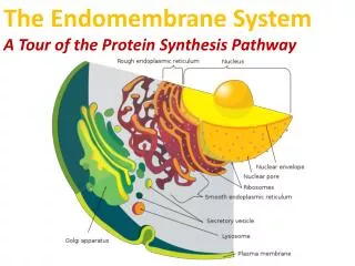
Image of nucleus, endoplasmic reticulum and Golgi apparatus.(1) Nucleus. (2) Nuclear pore. (3) Rough endoplasmic reticulum (RER). (4) Smooth endoplasmic reticulum (SER). (5) Ribosome on the rough ER. (6) Proteins that are transported. (7) Transport vesicle. (8) Golgi apparatus. (9) Cis face of the Golgi apparatus. (10) Trans face of the Golgi apparatus. (11) Cisternae of the Golgi apparatus.
2.2 The polarity of Golgi complex The Golgi complex is compartmentalized. Phosphorylation occurs in the Cis region. In other regions, different types of carbohydrates are added as a glycoprotein passes through the cisternae. This figure illustrates the different regions where sugars like mannose , galactose , etc are added. The final sorting is done in the Trans Golgi complex. So it is called the polarity organelle
2.3 The functions of Golgi complex 2.3.1 Glycosylation in the Golgi complex Golgi complex plays a key role in the assembly of the carbohydrate component of glycoproteins and glycolipids. O-linked oligosaccharides takes place in Golgi complex. Oligosaccharide chain islinked to the hydroxyl group of serine or threonine .
2.3.1 Glycosylation in the Golgi complex The important role of Glycosylation : 1.One might suspect that they function to aid folding and the transport process; for example,carbohydrate as a marker during protein folding in ER and the use of carbohydrate-binding lectins in guilding ER-to-Golgi transport. 2.Limit the approach of other macromolecules to the protein surface, more resist to digestion by proteases. 3.Regulatory roles in signaling through the cell-surface receptor notch, to allow these cells to respond selectively to activating stimuli.