CARBOHYDRATES: STRUCTURE AND FUNCTION
CARBOHYDRATES: STRUCTURE AND FUNCTION. Objectives. To understand the structure of carbohydrates of physiological significance To understand the main role of carbohydrates in providing and storing of energy To understand the structure and function of glycosaminoglycans. OVERVIEW.

CARBOHYDRATES: STRUCTURE AND FUNCTION
E N D
Presentation Transcript
Objectives To understand the structure of carbohydrates of physiological significance To understand the main role of carbohydrates in providing and storing of energy To understand the structure and function of glycosaminoglycans
OVERVIEW • The most abundant organic molecules in nature • provide important part of energy in diet • Act as the storage form of energy in the body • are structural component of cell membrane • The empiric formula is (CH2O)n – “hydrates of carbon”
OVERVIEW CONT’D • Diseases associated with disorders of carbohydrate metabolism: • Diabetes mellitus • Galactosemia • Glycogen storage diseases • Lactose intolerance
CLASSIFICATION: • Monosaccharides:Simple sugar • Disaccharides: 2 monosaccharide units • Oligosaccharides: 3-10 monosaccharide units • Polysaccharides:more than 10 sugar units Homopolysaccharides and heteropolysaccharides
Monosaccharides Further classified based on: 1. No. of carbon atoms 2. Functional group: Aldehyde group – aldoses Keto group – ketoses
Some Monosaccharides All carbons in a monosaccharide are bonded to a hydroxyl group (-OH) except for one which is bonded to a carbonyl group (=O) (note that this statement is true only for the linear form of monosaccharides)
Isomerism Isomers Compounds having same chemical formula but different structural formula The No. of isomers depends on the No. of asymmetric C
The two simplest sugars Some Monosaccharides Note Numerous Chiral Carbons
Sugar Isomers Aldo-keto Epimers D- and L-Forms α- and β-anomers
Aldo-Keto Isomers Example: Glucose and fructose
Epimers Epimers CHO dimers that differ in configuration around only one specific carbon atom -Glucose and galactose, C4 -Glucose and Mannose, C2 Galactose and mannose are not epimers, why?
Enantiomers (D- and L-Forms) Structures that are mirror imagesof each other and are designated as D- and L- sugars based on the position of –OH grp on the asymmetric carbon farthest from the carbonyl carbon Majority of sugars in humans are D-sugars
α- and β-Forms Cyclization of MonosaccharidesMonosaccharides with 5or more carbon are predominantly found in the ring form • The aldehyde or ketone grp reacts with the –OH grp on the same sugar • Cyclization creates an anomeric carbon (former carbonyl carbon) generating the α and β configurations
Mutarotation In solution, the cyclic α and β anomers of a sugar are in equilibrium with each other, and can be interconverted spontaneously Fischer Projection Haworth Projection
Disaccharides: Joining of 2 monosaccharidesby O-glycosidic bond: Maltose (α-1, 4) = glucose + glucose Sucrose (α-1,2) = glucose + fructose Lactose (β-1,4) = galactose + glucose Lactulose (β-1,4) = galactose + fructose
Disaccharides CONT’D Lactose
Polysaccharides Homopolysaccharides: Branched: glycogen ( storage in humans) and starch ( plants), both (α-glycosidic polymer of glucose) Unbranched: cellulose (β-glycosidic polymer) Heteropolysaccharides: e.g., glycosaminoglycans (GAGs)
Reducing Sugars If the O on the anomeric C of a sugar is not attached to any other structure, that sugar can act as a reducing agent Reducing sugars reduce chromogenic agents like Benedict’s reagent or Fehling’s solution to give a colored periceptate Urine is tested for the presence of reducing sugars using these colorimetric tests
Reducing Sugars CONT’D Examples: Monosaccharides Maltose and Lactose Sucrose is non-reducing, Why?
Complex Carbohydrates Carbohydrates attached to non-carbohydrate structures by glycosidic bonds (O- or N-type) e.g. 1. Purine and pyrimidine bases in nucleic acids 2. Aromatic rings in steroids 3. Proteins in glycoproteins and glycosaminoglycans 4. Lipids found in glycolipids 5. Bilirubin
Glycosidic Bonds • O-Glycosidic N-Glycosidic
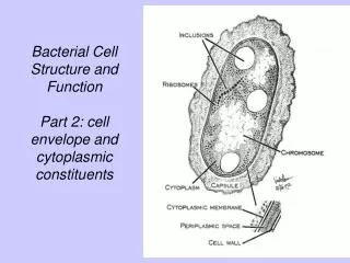
Glycosaminoglycans (GAGs) Glycosaminoglycans (GAGs) are large complexes of negatively charged heteropolysaccharide chains are associated with a small amount of protein, forming proteoglycans, which consist of over 95 percent carbohydrate bind with large amounts of water, producing the gel-like matrix that forms body's ground substance The viscous, lubricating properties of mucous secretions also result from GAGs, which led to the original naming of these compounds as mucopolysaccharides
Glycosaminoglycans (GAGs) GAGs are linear polymers of repeating disaccharideunits [acidic sugar-amino sugar] n The amino sugar (usually sulfated) is either D-glucosamine or D-galactosamine The acidic sugar is either D-glucuronic acid or L-iduronic acid GAGs are strongly negatively-charged: carboxyl groups of acidic sugars & Sulfate groups
Resilience of GAGs Being negatively charged GAG chains are extended in solution and repel each other and when brought together, they "slip" past each other This produces the "slippery" consistency of mucous secretions and synovial fluid When a solution of GAGs is compressed, the water is "squeezed out" and the GAGs are forced to occupy a smaller volume. When the compression is released, the GAGs spring back to their original, hydrated volume because of the repulsion of their negative charges This property contributes to the resilience of synovial fluid and the vitreous humor of the eye
Members of GAGs Examples of GAGs are: • Chondroitin sulfates • Keratan sulfates • Hyaluronic acid • Heparin
CHONDROITIN SULFATES Disaccharide unit: Sulfated N-acetyl-galactosamine +Glucuronic acid Mostabundant GAG in the body Form proteoglycan aggregates Found in cartilage, tendons, ligaments, and aortaIn cartilage, they bind collagen and hold fibers in a tight, strong network
KERATAN SULFATES Disaccharide unit: N-acetylglucosamine Galactose (no uronic acid) Sulfate content is variable and may be present on C-6 of either sugar Most heterogeneous GAGs Present in loose connective tissue and cornea
HYALURONIC ACID Disaccharide unit: N-acetylglucosamine Glucuronic acid Different from other GAGs: Unsulfated Not covalently attached to protein The only GAG found in bacteria Serves as a lubricant and shock absorber Found in synovial fluid of joints, vitreous humor of the eye, the umbilical cord, and cartilage
HEPARIN Disaccharide unit: Glucosamine and Glucuronic or iduronic acids Sulfate is found on glucosamine and uronic acid Unlike other GAGs that are extracellular, heparin is an intracellular component of mast cells that line arteries, especially liver, lungs and skin Serves as anticoagulant