NUCLEIC ACIDS: STRUCTURE and FUNCTION
NUCLEIC ACIDS: STRUCTURE and FUNCTION. NUCLEIC ACIDS: STRUCTURE and FUNCTION. Objectives: To describe the structure of DNA and RNA To explain how the structure of DNA relates to the functioning of the gene. NUCLEIC ACIDS: STRUCTURE and FUNCTION. Objectives:

NUCLEIC ACIDS: STRUCTURE and FUNCTION
E N D
Presentation Transcript
NUCLEIC ACIDS: STRUCTURE and FUNCTION
NUCLEIC ACIDS: STRUCTURE and FUNCTION • Objectives: • To describe the structure of DNA and RNA • To explain how the structure of DNA relates to the functioning of the gene
NUCLEIC ACIDS: STRUCTURE and FUNCTION Objectives: At the end of these lectures you should be able to describe: - The types of nucleic acids - The structure of nucleic acids - The base composition of DNA - The conformations of DNA - The functions of nucleic acids
NUCLEIC ACIDS: STRUCTURE and FUNCTION Why study nucleic acids? DNA is the focus of attention because of its role in carrying and expressing genetic information. The Human Genome Project where over 90% (99.9% accuracy) of the 3.2 billion nucleotideshave been cloned and sequenced. The information is hoped, will revolutionize the detection, prevention and treatment of conditions from cancer to depression to old age itself.
NUCLEIC ACIDS: STRUCTURE and FUNCTION In the foreseeable future, medical doctors will drip droplets of our genes onto a biochip to figure out if we have the kind of prostrate cancer that will kill or not. Scientist will learn which genes turn on when a wound heal. It is the letters ATCG… on and on for about 3.2 billion of such letters that provides the information.
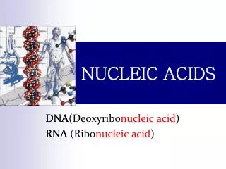
NUCLEIC ACIDS: STRUCTURE and FUNCTION In the eyes of the critic, it threatens to undermine privacy and brings on genetic discrimination in insurance and employment. The Structure of Nucleic Acids The chemistry of DNA has been studied since 1868 and by 1900 the basic chemistry of nucleic acids was worked out. By 1920, two forms of nucleic acids were differentiated:
NUCLEIC ACIDS: STRUCTURE and FUNCTION DNA and RNA Both deoxyribonucleic acid(DNA) and ribonucleic acid(RNA) are high-molecular-weight polymeric compounds. The chain-like macromolecule is made up of strings of monomeric units called nucleotides. On complete hydrolysis nucleic acids yields pyrimidine and purine bases, a sugar component and phosphoric acid.
NUCLEIC ACIDS: STRUCTURE and FUNCTION Each nucleotide is composed of three components: MONOMERIC COMPONENTS 1. Pentose and Deoxypentose sugar: a cyclic 5 carbon sugar
NUCLEIC ACIDS: STRUCTURE and FUNCTION • These sugars in polynucleotides occur in • - Either D-ribose in RNA or • 2'-deoxyribose in DNA • Ribose C2 = OH • Deoxyribose C2 = H
NUCLEIC ACIDS: STRUCTURE and FUNCTION 2. The Nitrogenous bases, which is either Pyrimidine or a Purine derivative. The bases are planar, aromatic, heterocyclic molecules. Purine Bases
NUCLEIC ACIDS: STRUCTURE and FUNCTION Pyrimidine bases The pyrimidine bases are derivatives of the parent compound pyrimidine and includes = thymine,cytosine and uracil.
NUCLEIC ACIDS: STRUCTURE and FUNCTION The bases, Adenine, Guanine and Cytosine are found in both DNA and RNA. Thymine is present only in DNA. Uracil is present only in RNA. In certain of the bacterial viruses cytosine is replaced by 5-methylcytosine or 5-hydroxymethylcytosine. Both the purine and pyrimidine bases can undergo keto-enol tautomerism
NUCLEIC ACIDS: STRUCTURE and FUNCTION 3. Phosphate group A molecule of Phosphoric acid, PO43- Nucleosides When a purine or a pyrimidine base is linked to ribose or deoxyribose the resulting compound is known as a nucleoside. The nucleosides from ribose = ribonucleosides The nucleosides from 2-deoxyribose = deoxyribonucleosides
NUCLEIC ACIDS: STRUCTURE and FUNCTION The nucleosides derived from 2-deoxyribose are known as: deoxyadenosine, deoxyguanosine, deoxycytidine, deoxythymidine etc.
NUCLEIC ACIDS: STRUCTURE and FUNCTION Nucleotides When a purine or a pyrimidine is linked to ribose or deoxyribose and phosphoric acid the resulting compound is known as a nucleotide. Those derived from ribonucleosides are referred to as ribonucleotides. Those derived from deoxyribonucleosides are referred to as deoxyribonucleotides.
NUCLEIC ACIDS: STRUCTURE and FUNCTION Nucleotides Since the ribonucleosides have three free hydroxyl groups on their sugar ring, three possible monophosphate can be formed. The ribo and deoxyribo nucleoside 5'-phosphate may be further phosphorylated at position 5' to yield 5'-di- and 5'-tri-phosphates. e.g. Adenosine 5'-phosphate (AMP), Adenosine 5'-diphosphate (ADP), Adenosine 5'-triphosphate (ATP)
NUCLEIC ACIDS: STRUCTURE and FUNCTION The Primary Structure of Nucleic Acids In a nucleotide, a base is attached to a pentose sugar by N-glycosidic bonds to carbon # 1 of the sugar and a nitrogen atom of the base. Sugar is attached at position N-1 of the pyrimidine base. Sugar is attached at position N-9 of the purine base.
NUCLEIC ACIDS: STRUCTURE and FUNCTION The Primary Structure of Nucleic Acids The phosphate is attached to the 5' carbon of the sugar by phosphodiester linkages. The phosphate is responsible for the strong negative charge of nucleic acids. Nucleic acids are polyanions. Chemically, nucleic acids are composed of covalently linked chains of nucleotides.
NUCLEIC ACIDS: STRUCTURE and FUNCTION The Primary Structure of Nucleic Acids Linkage between 5'-Phosphate and 3'-OH group. 3'-5'-internucleotide linkage in both DNA and RNA was confirmed by Cohn and his colleagues in 1956.
NUCLEIC ACIDS: STRUCTURE and FUNCTION DNA base composition In 1952, Chargaff described fundamental features of DNA: The sum of purines is equal to the sum of pyrimidines. The sum of the amino bases is equal to the sum of keto bases. This equivalence of A and T, and G and C are importance in relation to the formation of the DNA double helix.
NUCLEIC ACIDS: STRUCTURE and FUNCTION DNA base composition cont’d DNA isolated from different species reveals wide variations in the molar proportions of bases. This is independent of the age of the organism, its nutritional state or any environmental factor. The ratio, A+T/G+C, called the base ratiomay vary widely between species, and remains constant for any one species. These relationships are referred to as Chargaff's rule
NUCLEIC ACIDS: STRUCTURE and FUNCTION Molar proportion of bases (as moles of base per 100 moles of phosphate) in DNAs from various sources.
NUCLEIC ACIDS: STRUCTURE and FUNCTION DNA base composition cont’d DNA's base composition varies widely among different organism G + C ranges from 25 - 75% in different species of bacteria; this is relatively constant among related species. G + C ranges from 39 - 46% in mammals. With RNA these rules do not apply - ss RNA With ds RNA in certain viruses the rule would apply
NUCLEIC ACIDS: STRUCTURE and FUNCTION Molecular Weight of DNA The molecular weight of DNA ranges from 106 to 1010 daltons. The size of the DNA molecules is now customarily described in terms of Kilobase pairs (kb) rather than Daltons or Molecular weight. A molecular weight of one million corresponds approx. to 1.5 kb. E.g. E. coli MW 3 x 10⁹ ~ 4500 kb.
NUCLEIC ACIDS: STRUCTURE and FUNCTION The Secondary Structure of DNA James Watson and Francis Crick published in Nature the 3-dimensional structure of the DNA molecule. The impact of their model has been enormous and it constitutes as the foundation of modern molecular biology. Ref. Watson, J.D. and Crick, F.H.C. (1953) Nature, 171, 373 Ref. Franklin, R. and Gosling, R. G. (1953) Nature, 171, 740; 172, 156. (Crick died Aug. 2004: He was 88 years old)
NUCLEIC ACIDS: STRUCTURE and FUNCTION The Secondary Structure of DNA The study of the molecular architecture started long before Watson and Crick, actually by Astbury (1947), and later by Rosalind Franklin and Gosling (1953). X-ray diffraction was used to elucidate the secondary structure of DNA. The diffraction pattern of the mounted fibre is then recorded in an atmosphere of controlled humidity.
NUCLEIC ACIDS: STRUCTURE and FUNCTION • The Secondary Structure of DNA • Watson and Crick in 1953 proposed that the DNA molecule extended chain having a highly ordered structure and is composed of: • two complementary polymeric chains twisted about each other. • the two stands run in opposite directions (antiparallel alpha-helices), and are of opposite polarity.
NUCLEIC ACIDS: STRUCTURE and FUNCTION • The Secondary Structure of DNA • the rails of the ladder run in opposite direction contain alternating units of deoxyribose sugar and phosphate. • the polynucleotide chain, the sugar and phosphate groups are always linked together by 3’ - 5’ phosphodiester linkages. • the purine and pyrimidine bases are flat (planar), are relatively water-insoluble and are stack tightly on top of one another like a pile of plates.
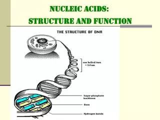
NUCLEIC ACIDS: STRUCTURE and FUNCTION • The Secondary Structure of DNA • the bases are arranged at right angles to the long axis of the polynucleotide chain. • each step is composed of a pair of nucleotides. • the order of the purine and pyrimidine bases along the chain is highly irregular. • the chain is not straight but is wound helically around a central axis, one full turn ( the pitch) of the helix extending 3.4 nm (34 Å), and there are 10 bases per turn.
NUCLEIC ACIDS: STRUCTURE and FUNCTION • The Secondary Structure of DNA • the bases are separated by a spacing of 0.34 nm (3.4 Å). • the width of the double helix is approx. 2 nm (20 Å). • the chains are complementary, in that the sequence of bases on one strand is the exact complement of the other strand.
NUCLEIC ACIDS: STRUCTURE and FUNCTION The Secondary Structure of DNA This form of the double helix is known as the B-form. Complementary strand of the opposite polarity lying below: 5'-ACGT-3' 3'-TGCA-5‘
NUCLEIC ACIDS: STRUCTURE and FUNCTION The Secondary Structure of DNA
NUCLEIC ACIDS: STRUCTURE and FUNCTION • The Secondary Structure of DNA • There are two reasons why the bases must pair in this specific way: • The purine, with a double ring are larger structures than pyrimidine, with a single ring. If two purine are paired their dimensions are too great to fit the constant diameter of the double helix (2 nm) while the dimensions of the two pyrimidine are too small.
NUCLEIC ACIDS: STRUCTURE and FUNCTION The Secondary Structure of DNA 2.The second determinant of specificity is the positions on the bases of the hydrogen atoms that can participate in bonding. This is essential to the biological functioning of DNA. If the hydrogen atoms had no fixed positions, ade could often pair with cytosine and gua with thymine. The complementary relationship between the opposing chain sequences that gives DNA the capacity for self-replication.
NUCLEIC ACIDS: STRUCTURE and FUNCTION The Secondary Structure of DNA Occasionally, H-atoms do undergo shift to other positions and when this occurs new pairing interactions may be possible, this is called tautomeric shifts. The nitrogen atoms attached to the purine and pyrimidine rings are usually in the amino (NH2) form and only rarely assume the imino (NH) configuration,
NUCLEIC ACIDS: STRUCTURE and FUNCTION The Secondary Structure of DNA Likewise, the oxygen atoms attached to C6 atoms of guanine and thymine normally have the keto (C-O)form and rarely take up the enol (COH) configuration. If the H-atom normally present at the 6-amino position in adenine shifts to the N1 position, Ade will pair with cytosine instead of with thymine.
NUCLEIC ACIDS: STRUCTURE and FUNCTION • DNA Conformations • Wilkins and his colleagues demonstrated that, depending on the conditions chosen to produce the DNA fibres, they can have a variety of possible conformations (structures). • The major forms are the: • B-form, basically describes the Watson and Crick model, • A-form DNA, • Z-form DNA.
NUCLEIC ACIDS: STRUCTURE and FUNCTION DNA Conformations:Structural features of DNA
NUCLEIC ACIDS: STRUCTURE and FUNCTION DNA Conformations : B-form DNA B-form DNA is thought to represent the conformation of most DNA found in cells. The main features that distinguish B-form DNA from other forms are: the pitch, the angle of tilt that the base pairs make with the helical axis, and the distinct major and minor grooves. The B-DNA is long and thin.
NUCLEIC ACIDS: STRUCTURE and FUNCTION DNA Conformations : B-form DNA There is a lot of water associated with DNA. Up to 72 molecules per 12 base pairs. The hydrogen bonding potential is much greater in the major groove than in the minor groove and there appears to be greater dependence on base sequence for interaction as it was later shown that proteins attach to sequence-specific DNA segments.
NUCLEIC ACIDS: STRUCTURE and FUNCTION DNA Conformations : B-form DNA
NUCLEIC ACIDS: STRUCTURE and FUNCTION DNA Conformations : A-form DNA The B-DNA is converted to A-DNA by tilting the base pairs some 30 deg so that successive base pairs occur every 0.28 nm.
NUCLEIC ACIDS: STRUCTURE and FUNCTION DNA Conformations : A-form DNA The B-DNA is converted to A-DNA by tilting the base pairs some 30 deg so that successive base pairs occur every 0.28 nm. The bases are tilted and lie well off the axis as a result the A-DNA is short and broad as with deeper and narrow major grooves. This is the predominant conformation of DNA-RNA hybrids and double stranded RNA segments.
NUCLEIC ACIDS: STRUCTURE and FUNCTION DNA Conformations : Z-form DNA DNA containing alternating purine and pyrimidine residues (dinucleotides: CGCGCGCG) can fold up into left-handed as well as right handed helices. The phosphate in the backbone is zig-zagged due to the fact that the repeating unit is a dinucleotide rather than a mononucleotide – hence the name Z-DNA. Z-forms will also form in 1 mM MgCl2 if the C5 of cytosine is substituted with methyl, bromo or iodo groups.
NUCLEIC ACIDS: STRUCTURE and FUNCTION DNA Conformations : Z-form DNA Z-DNA contains 12 bases per turn, and a pitch of 4.5 nm, and inclination of base pair from the horizontal ~9°, this gives the overall Z-DNA an elongated and slimmer look. Only very minor amounts of Z-DNA exists in-vivo, and is probably important immunologically.
NUCLEIC ACIDS: STRUCTURE and FUNCTION DNA Conformations : Z-form DNA
NUCLEIC ACIDS: STRUCTURE and FUNCTION Deformation of the double helix The sugar phosphate backbone causes the double helix to be quite rigid. Also important in conferring rigidity is the stacking of the bases.
NUCLEIC ACIDS: STRUCTURE and FUNCTION Deformation of the double helix In solution DNA is quite a plastic molecule it is constantly subjected to localized thermally induced fluctuations in the arrangement of its atoms which causes individual molecules to bend, twist and stretch. DNA that tends to kink reveals a set of four CAAAAAT or CAAAAAAT segments and was found to be separated by single turns (10-nt) of the double helix.
NUCLEIC ACIDS: STRUCTURE and FUNCTION conformation DNA can either be linear or circular. Most if not all bacterial chromosomes are circular. Certain phages or viruses have linear DNA e.g. Lamda phage, adenovirus, poxvirus. Some molecules that are linear when isolated from a virus particle are found as circular forms inside the host.
NUCLEIC ACIDS: STRUCTURE and FUNCTION conformation DNA is naturally supercoiled and is biologically very important. Supercoiled refers to the twisting of the double helical DNA. DNA is naturally negatively supercoiled. DNA can be negatively suprecoiled (right handed) or positively supercoiled (left handed). Negative Supercoiling results from under-winding or unwinding, where as positive supercoiling results from tighter winding.