Download
1 / 10
100 likes | 390 Views
Vision & Alzheimer ’ s Disease. Victoria S. Pelak , MD University of Colorado School of Medicine March 2014. Alzheimer ’ s Disease (AD). AD affects over 5 million people in the United States alone and accounts for 50-90% of all cases of dementia.

E N D
Vision & Alzheimer’s Disease Victoria S. Pelak, MD University of Colorado School of Medicine March 2014
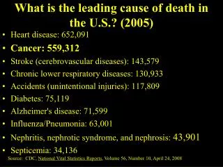
Alzheimer’s Disease (AD) • AD affects over 5 million people in the United States alone and accounts for 50-90% of all cases of dementia. • It is an age-related neurodegenerative disorder with progressive loss of cognitive function over time. • AD is a pathologic diagnosis made at autopsy with pathological changes of intracellular tangles and extracellular plaques. • A clinical diagnosis for Probable AD Dementia requires the following: a loss of cognitive function in two or more cognitive domains (or in one cognitive domain along with a change in personality or behavior or comportment), insidious progression, and inability to function in daily activities. • Patients with AD have visual symptoms that are often misdiagnosed or overlooked.
Alzheimer’s Disease and Vision • Posterior regions of the brain process visual information and are greatly affected by the pathological changes and synaptic loss due to AD. • Retinal ganglion cells and their axons (retinal nerve fibers) degenerate in AD but do not cause the classic clinical syndrome of an optic neuropathy. • Vision complaints can be the first sign of AD in up to 1/3 patients, and patients will often seek help from eye providers for 1-5 years before the recognition of AD.
Functional Anatomy of the Brain The occipital lobe is the hub of the visual cortical network – with billions of neurons – and it distributes information based on content
Two Visual Processing Streams:The What and Where Pathways • In AD, the where pathway (or dorsal stream) is typically affected earlier, and to a greater extent, than the what pathway (or ventral stream). • Visuospatial dysfunction manifests when the where pathway is disrupted. • Difficulty recognizing colors, faces, or objects occurs when the what pathway is disrupted. • Area V5/MT is also affected by AD, and it is important for motion processing during way finding and driving. Visual areas of the cortex have a hierarchy and are sequentially labeled with area V1 as the primary visual area (the striate cortex in humans).
AD: Progression of Visual Brain Problems • Early stages, patients might have: • Trouble reading books or newspapers • Trouble driving • Mid-stages, patients might: • Stop reading • Be unable to drive • Start to get lost • Late stages, patients might: • Wander • Be unable to recognize a familiar face or objects (forms of agnosia) • Be unable to reach out for objects within their sight (optic ataxia) • Be unable to move their eyes to a visual target (ocular motor apraxia) • Be unable to recognize a visual scene (unable to use the parts of the scene to form a precept of the scene as a whole – simultanagnosia)
Variations on Presentation of AD • Visual Variant of AD: • Visual symptoms are more prominent than memory symptoms at onset and throughout the disease process. • Posterior Cortical Atrophy or PCA: • Visual symptoms dominate and other areas of cognition not affected or only minimally affected at onset. • Most patients have a best correct visual acuity of 20/20 in both eyes at the time of their first visual complaint, and they develop progressive cortical vision loss resulting in decreased visual acuity over time. • Most patients will progress to Alzheimer’s disease.
PCA: In their own wordsBelow is a list, provided by the family, of symptoms experienced by a patient with PCA
Associated Features of PCA • Apraxia (constructional, dressing, limb, ideomotor) • Language: • Alexia • Agraphia • Transcortical sensory aphasia • Note: visual hallucinations occur in a minority of patients with PCA and depend on the pathological cause of PCA (i.e. more common with Lewy Bodies than with Alzheimer’s pathology)
PCA with Autopsy - 37 Total Authors: Renner, Tang-Wai, Victoroff, Ala, Schmidtke, Pelak AD=Alzheimer’s Disease LBD=Lewy Body Disease pathology PD=Parkinson’s Disease CBD=Cortical Basal Degeneration CJD=Creutzfeldt-Jakob Disease FFI=Fatal Familial Insomnia DLB=Dementia with Lewy Bodies