Download
1 / 168
1.68k likes | 2.13k Views
10. The Nervous System: Part Two: The Traffic Control Center. Multimedia Asset Directory. Slide 23 PET Scan Animation Slide 77 Parkinson's Disease Video Slide 128 Absence Seizures Video Slide 129 Alzheimer's Disease Video Slide 130 Autism Video Slide 131 Bipolar Disorder Video

E N D
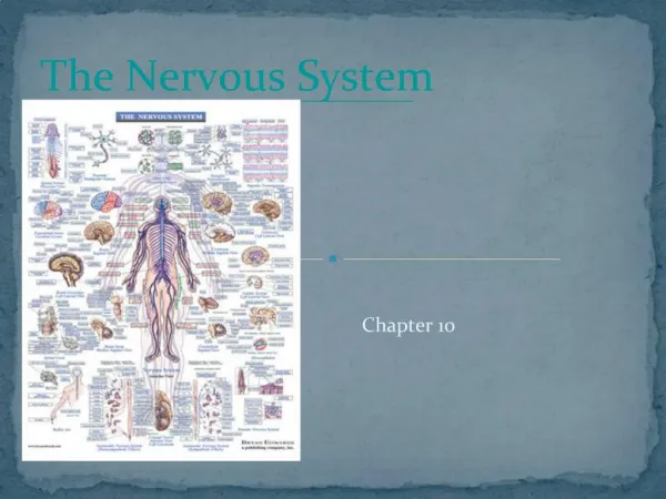
10 The Nervous System: Part Two: The Traffic Control Center
Multimedia Asset Directory Slide 23 PET Scan Animation Slide 77 Parkinson's Disease Video Slide 128 Absence Seizures Video Slide 129 Alzheimer's Disease Video Slide 130 Autism Video Slide 131 Bipolar Disorder Video Slide 132 Dissociative Identity Disorder Video Slide 133 Epilepsy Video Slide 134 Obsessive Compulsive Disorder Video Slide 135 Schizophrenia Video Slide 136 Complex Partial Seizures Video Slide 137 Generalized Tonic-Clonic Seizure Video
Multimedia Asset Directory Slide 138 Panic Attacks Video Slide 139 Delirium Animation Slide 140 Stroke Animation Slide 141 Shock Animation Slide 142 Pharmacy Video
Introduction • In this chapter we will focus on the main control of the nervous system – the brain. • Then, we will put the whole system together to show the big picture of how the nervous system functions.
Learning Objectives • Organize the hierarchy of the nervous system. • Locate and define the internal and external structures and their corresponding functions of the brain. • List and describe the cranial nerves and their functions.
Learning Objectives • Describe the sensory and motor functions of the brain with related structures. • Contrast the parasympathetic and sympathetic branches of the autonomic nervous system. • Discuss some representative diseases of the nervous system.
anterior commissure (an TEE ree or KAHM ih shoorz) basal nuclei (BAY sal noo KLEE eye) cerebellum (ser eh BELL um) cerebrum (ser EE brum) corpus callosum (KOR pus kah LOH sum) diencephalon (DYE in SEFF ah lon) fornix (FOR niks) gyri (JIE rie) hypothalamus (high poh THAL ah mus) Pronunciation Guide Click on the megaphone icon before each item to hear the pronunciation.
limbic system (LIM bick) medulla oblongata (meh DULL ah OB long GA ta) occipital lobe (awk SIP eh tal) parietal lobe (pah RYE eh tal) pineal body (PIN ee al) subarachnoid space (sub ah RACK noyd) sulcus (SULL cus) thalamus (THAL ah mus) Pronunciation Guide Click on the megaphone icon before each item to hear the pronunciation.
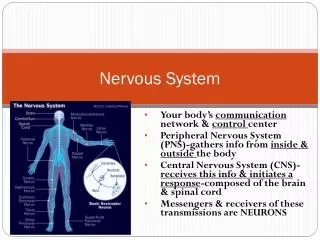
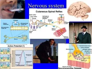
The Brain and Cranial Nerves • The brain and cranial nerves represent the major controls of the nervous system. • The brain acts more as the main processor and director of the entire system. • The cranial nerves leave the brain and go mainly to the head where they receive information and send it back to the brain (sensory) and the brain sends back instructions to move (motor).
The Brain • At the top of the spinal cord, beginning at the level of the foramen magnum and filling the skull, is the brain. • The brain can be divided into several anatomical and functional sections. • External anatomy • Cerebrum • Cerebellum • Brain stem
Cerebrum • The cerebrum is the largest part of the brain. • It is divided into the right and left hemisphere by the longitudinal fissure and divided from the cerebellum by the transverse fissure. • The surface of the cerebrum is not smooth, but broken by ridges (gyri) and grooves (sulci) collectively known as convolutions.
Cerebrum • These convolutions serve a very important purpose by increasing the surface area of the brain, so you can pack more brain in a smaller space. • Most of the sulci are extremely variable in their locations among humans, but a few are in basically the same place in every brain. These divide the brain into lobes.
Lobes of the Brain • The lobes are named for the skull bones that cover them and occur in pairs, one in each hemisphere. • The most anterior lobes, separated from the rest of the brain by the central sulci are the frontal lobes. The frontal lobes are responsible for motor activities, conscious thought, and speech.
Lobes of the Brain • Posterior to the frontal lobe are the parietal lobes. The parietal lobes are involved with body sense perception and language comprehension. • Posterior to the parietal lobes are the occipital lobes, which are responsible for vision. • The most inferior lobes, separated by the lateral sulci, are the temporal lobes, which are involved in hearing and integration of emotions.
Lobes of the Brain • There is a section of the brain, the insula, deep inside the temporal lobes that is often listed as the fifth lobe, but is not visible on the surface of the cerebrum. • Much of the information coming into your brain is contralateral, meaning that the right side of your body is controlled by the left side of your cerebral cortex and the left side of your body is controlled by the right side of your cerebral cortex.
Specific Regions of Cerebrum • On either side of the central sulcus are two gyri named for their locations: the precentral gyrus, anterior to the central sulcus, and the postcentral gyrus, posterior to the central sulcus. • The frontal lobe also contains Broca’s area, which controls motor output for speech.
Specific Regions of Cerebrum • In the parietal lobe is Wernicke’s area. Wernicke’s area was long thought to control sensory aspects of language, including understanding. This area is a general interpretive area for many types of sensory information and may integrate much of the sensory information coming to the cerebral cortex. • In most people, Broca’s and Wernicke’s areas are in the left hemisphere.
The Cerebellum • The cerebellum is posterior to the cerebrum. • It too is divided into hemispheres by a raised ridge called the vermis. • The surface is convoluted like that of the cerebrum. • From its external appearance it is easy to see why the cerebellum is called the little brain. • The cerebellum is involved in sensory and motor coordination and balance.
Click here to view a video on the topic of PET Scans. Back to Directory
The Brain Stem • The brain stem is a stalk-like structure inferior to, and partially covered by, the cerebrum. • The brain stem is divided into three sections. • The medulla oblongata is continuous with the spinal cord. Responsible for control of heartbeat, respiration, and blood vessel diameter.
The Brain Stem • The brain stem is divided into three sections. • The pons is just superior to the medulla oblongata and plays a role in respiration. • The midbrain is the most superior portion of the brain stem and is completely covered by the cerebrum. The midbrain is a pathway to relay visual and auditory impulses and other information to the cerebrum.
The Brain Stem • The brain stem receives sensory information and contains control systems for vital processes such as blood pressure, heart rate, and ventilation.
From the Streets:Nervous System Infections • Infections of the nervous system can be life-threatening. • Meningitis • The most common NS infection. • Infection of the meninges • Types • Bacterial • Viral • Fungi • Parasites • Prions
From the Streets:Nervous System Infections • Meningitis • Causes • Signs and symptoms • Brudinski’s sign • Kernig’s sign • Diagnostic tests • Treatment
Internal Anatomy of the Brain • The inside of the brain has white and gray matter, along with hollow cavities containing CSF. • The white matter of the brain is surrounded by the gray matter. • The layer of gray matter surrounding the white matter is called the cortex. In the cerebrum it is called the cerebral cortex and in the cerebellum it is called the cerebellar cortex. • Deep islands of gray matter are nuclei.
Internal Anatomy of the Brain • Ventricles – the cavities in the brain. They are continuous with the central canal of the spinal cord and the subarachnoid space of both the brain and the spinal cord. • The lateral ventricles (ventricle 1 and 2) are in the cerebrum, the third ventricle is in the diencephalon (a region between the cerebrum and brain stem) and the fourth ventricle is in the inferior part of the brain between the medulla oblongata and the cerebellum.
From the Streets:Burr Holes • An epidural hematoma may result from head trauma causing bleeding in the epidural space. • This causes rapid swelling and compression of the brain. • A physician may drill burr holes over the hematoma to relieve pressure.
Figure 10-3 (continued) (B) Sagittal sectional view of the brain.
Figure 10-3 (continued) (C) Frontal sectional view of the brain.
Clinical Application: CSF Circulation and Hydrocephalus • The ventricles of the brain, the central canal of the spinal cord, and the subarachnoid space surrounding both the brain and spinal cord are filled with CSF. The CSF is filtered from blood in the ventricles by tissue called choroid plexus.
Clinical Application: CSF Circulation and Hydrocephalus • CSF made in the lateral ventricles flows through a tiny opening into the third ventricle and then through another opening into the fourth ventricle. CSF flows into the central canal of the spinal cord and the subarachnoid space. CSF is returned to the blood via special “ports” between subarachnoid space and blood spaces in the dura mater.
Clinical Application: CSF Circulation and Hydrocephalus • The balance of CSF made and CSF reabsorbed by the blood is very important. The brain is a very delicate organ captured between the liquid CSF and the bones of the skull. If there is too much CSF, pressure inside the skull will rise and eventually crush brain tissue.
Clinical Application: CSF Circulation and Hydrocephalus • This condition, in which there is too much CSF, is called hydrocephalus (water on the brain). Hydrocephalus can be caused by blockage of the narrow passages due to trauma, a birth defect, tumor, or decreased reabsorption of CSF. It can be treated by medication or, more commonly, a shunt is surgically placed to drain fluid to the heart or abdominal cavity.
Figure 10-4 Typical appearance of a infant with hydrocephalus.
The Cerebrum • The inside of the cerebrum reflects the external anatomy. The lobes (frontal, parietal, temporal, and occipital) are clearly visible. • The right and left hemispheres are connected by several white matter pathways surrounding the lateral ventricles called the corpus callosum, the fornix, and the anterior commissure.
The Cerebrum • Several prominent cerebral nuclei are also involved in motor coordination, sensation, and motor control.
The Diencephalon • Inferior to the cerebrum is a section of the brain, that is not visible from the exterior, called the diencephalon. • Consists of several parts including the thalamus, hypothalamus, pineal body, and the pituitary gland which interface with the endocrine system.
The Diencephalon • The diencephalon contains the third ventricle and a number of nuclei. • Basal nuclei and limbic system • Nuclei responsible for controlling hormone levels, hunger and thirst, body temperature, sleep-wake cycles, and for coordination of the flow of information around the brain
The Cerebellum • The external similarities between the cerebellum and cerebrum are also obvious internally. • The cerebellum has a gray matter cortex and a white matter center, known as the arbor vitae (tree of life). • The cerebellum also has nuclei that coordinate motor and sensory activity. • Essentially, the cerebellum fine tunes voluntary skeletal muscle activity and helps in the maintenance of balance.
Cranial Nerves • In order for the CNS to function, it must be connected to the outside world via nerves of the PNS. • Like the spinal cord has spinal nerves the brain has nerves called cranial nerves. • Cranial nerves are like spinal nerves in that they are the input and output pathways for the brain. • There are only 12 pairs of cranial nerves, all but two of which arise from the brain stem.
Cranial Nerves • Cranial nerves are not all mixed nerves like the spinal nerves. Some are mainly sensory and others are mainly motor, and some are mixed nerves. • Cranial nerves are much more specialized than spinal nerves. • Cranial nerves carry sensory and motor information for the head, face, and neck, as well as visual, auditory, smell, or taste sensations.