Download
1 / 43
2.07k likes | 6.43k Views
NURS 347 Towson University. Musculoskeletal Assessment. Musculoskeletal Assessment Fundamentals:. Anatomy and Physiology. Bones: 206 126 appendicular, 80 axial Joints: Where two or more bones join Muscles: Contraction = movement Voluntary skeletal muscles under conscious control

E N D
NURS 347Towson University Musculoskeletal Assessment
Musculoskeletal Assessment Fundamentals: Anatomy and Physiology
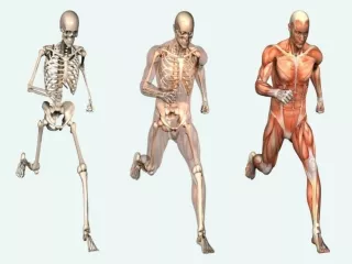
Bones: 206 126 appendicular, 80 axial • Joints: Where two or more bones join • Muscles: Contraction = movement • Voluntary skeletal muscles under conscious control • Support: Maintain stature • Movement • Protect vital organs • Produce red blood cells in bone marrow (hematopoiesis) • Storage of minerals, such as calcium and phosphorus Structure & Function
Synovial Joints • Bones are separated but enclosed in a joint cavity • Opposing bones covered with cartilage • Freely moveable • Ligaments:Fibrous bands connect two bones, strengthen joint • Bursa:Enclosed sac filled with synovial fluid that aim to reduce friction in areas such as the knee, shoulder. • Tendon:Attached the skeletal muscle to the bone Joints (Articulation)
Active and Passive Range of Motions should the be same • Active: When the patient can perform range of motion independently • Passive: When the patient has a limitation • Anchor the joint with one hand • Use your other hand and move to the joint’s limit Range of Motion
Assessing and Documenting a Limitation: • Goniometer: Used to precisely measure joint angles Range of Motion
Flexion: Bending limb at a joint Extension: Straightening a limb at a joint Range of Motion
Abduction: Moving a limb away from body’s midline Adduction: Moving a limb towards the body’s midline Range of Motion, continued
Pronation: Turning forearm so palm is down Supination: Rotating forearm so palm is up Range of Motion, Continued
Internal Rotation: External Rotation Range of Motion, Continued
Inversion: Moving the sole of the foot inward at the ankle Eversion: Moving the sole of the foot outward at the ankle Range of motion, Continued
Circumduction: Movement of the arm in a circle around the shoulder Range of Motion, Continued
Joints:Pain, stiffness, swelling, warmth, or limited range of movement? Muscles: Cramps, pain, or weakness? Bones: Pain, deformity, trauma (fractures, sprains, dislocations?) Activities of Daily Living:Any difficulty bathing, toileting, dressing, eating, communicating, or mobility? Occupational Hazards:Heavy lifting, repetitive movement? Self-Care:Recent weight gain, exercise program? Subjective Interview
Skeleton: Symmetry of skeleton; Size and contour of joint(s) Skin: Color or swelling Gait: Steady or unsteady Inspection
Joints Muscles Bones Range of Motion Tenderness Crepitus Muscle Strength Palpation
Inspect paired joints for: • Symmetry • Size • Contour • Color • Swelling • Deformities or Masses Joints: Inspection
Palpate to: • Stage edema: Pitting versus Non-Pitting • Masses • Warmth • Tenderness • Range of Motion (ROM) • Crepitus: Joints: Palpation An audible or palpable “crunching” or “grating” with movement
Head to Toe Musculoskeletal Assessment
Inspect: Area anterior to ear for: • masses, symmetry, discoloration • Palpate: • Crepitus or tenderness • temporalis and masseter muscles when teeth are clenched • Range of Motion: • Open mouth maximally Vertical motion • Partial mouth open Lateral motion • Stick outlower jaw Protrusion without deviation Temporomandibular Joint
Inspect the alignment of the head and neck • Palpate the spinous processes and sternomastoid, trapezius, and paravertebral muscles • Range of motion: • Chin to chest 45’ flexion • Chin to ceiling 55’ hyperextension • Touch ear to shoulder 40’ lateral bend • Turn chin to shoulder 70’ rotation DO NOT ASSESS IF SUSPECTED CERVICAL TRAUMA Cervical Spine
Inspect posteriorly and anteriorly: • Joint size and contour • Equality of bony landmarks • Palpate: • Spasm • Atrophy • Swelling • Heat • Tenderness • Crepitus during ROM Upper Extremities: Shoulder
How would you assess Range of Motion? Upper extremities: Shoulders
How would you assess Range of Motion? • Circumduction • Abduction • Adduction • Internal Rotation • External Rotation Upper extremities: Shoulders
Strength • Shrug Shoulders (also assesses which CN?) • Flex arms forward and up against resistance Upper extremities: Shoulders
Inspect joint and tissue • Range of Motion: • Bend and Straighten elbow (Flexion and Extension) • With slightly extended elbow, touch thefront and back of the hand to the table (Pronation and Supination) • Strength: Flex and extend elbow against resistance Upper Extremities: Elbow
Inspect joints (knuckles) and surrounding skin Palpate for warmth, crepitus, tenderness, or nodules Upper Extremities: Wrist and Hands
ROM: • Bend hand up and down at wrist • Bend fingers at metacarpophalangeal joints • Palms flat on table: Rotate in and outward • Spread fingers apart, make a fist • Touch thumb to each finger Upper Extremities: Wrist and Hands
Inspection of the Hips should be delayed until spinal he assessment With patient in the supine position, palpate the hip joints for crepitus or tenderness Lower Extremities: Hip
Range of Motion • Raise each leg with knee extended • Bend each knee up to the chest, keeping the other leg straight • Extend leg straight, then direct foot inward and outward • Swing leg laterally and medially, keeping knee straight Lower Extremities: Hip
Inspection and Palpation: • Skin free from lesions, smooth and even in coloring • Bilateral comparison: length and alignment • Swelling or fullness at the knee, pre- and suprapatellar bursa • Atrophy at quadriceps • Strength: Ask patient to push your hand away using their foot, assessing quadriceps’ strength Lower Extremities: Knee
Range of Motion • Bend and Extend each knee • Assess ROM during ambulation Lower Extremities: Knee
Inspect and compare both feet and toes, and their position. Examine: • skin color • Lesions • Contour • alignment with the upper leg • Note areas of calluses or bursal reactions, as they reveal areas of abnormal friction Lower Extremities: Foot and Ankle
Range of Motion • Point toes towards floor • Point toes towards nose • Turn soles of feet in and out • Flex and straighten toes • Strength • Maintain dorsiflexion and plantar flexion against resistance (hand) Lower Extremities: Foot and Ankle
Stand behind patient so you can see the entire back Inspect for spine’s straightness by following an imaginary vertical line from the head to the gluteal cleft Inspect for symmetry of shoulders, scapulae, and iliac crests (hips) bilaterally. Knees should be aligned and pointing forward Spinal Assessment: Posterior
Inspect for normal curvature of the spine • Convex thoracic curve • Concave lumbar curve • Range of Motion • Bend forward, touch toes • (concave curve should disappear) • Bend Sideways (35’) • Bend backward (Hyperextension 30’) • Assess for pain and decreased ROM Spinal Assessment: Lateral
Infants • Barlow-Ortolani’sManeuver: • Assesses for congenital dislocation of hips in infants. • Normal finding reveals smooth abduction and adduction of bilateral legs while in the supine position • SpinaBifida: • A tuft of hair over a dimple on the spinal midline may indicate spinal bifida • Children • Juvenile Rheumatoid Arthritis: Discomfort greater • in the morning, decreased ROM and pain in bilateral joints. • Scoliosis: Spinal asymmetry • Adolescents • Scoliosis: Spinal asymmetry Age Considerations: Infants & Children
Older Adults • Dorsal kyphosis • Rheumatoid Arthritis: Bilateral joint pain and decreased ROM, worse in the morning • Osteoarthritis: Unilateral or unrelated joint pain in which pain increases later in the day • Osteoporosis: Risk Factors & Prevention • Pregnancy • Waddling Gait • Backache • Muscle cramps • Lordosis Age Considerations & Pregnancy
Walk (with shoes on) to observe gait and balance Climb up and down stairs to assess balance and bilateral strength Pick up object from the floor Rise from sitting in chair Rise from lying in bed Functional Assessment