Download
1 / 32
320 likes | 477 Views
Linear dichroism for the study of nucleci acids, fibrous and membrane proteins. Electrons bonds structure. UV/visible light: l ~ 180 nm – 800 nm, energy h n = hc/ l causes electrons to go to higher energy levels. l required depends on electron rearrangement needed.

E N D
Linear dichroism for the study of nucleci acids, fibrous and membrane proteins
Electrons bonds structure UV/visible light:l ~ 180 nm – 800 nm, energy hn = hc/l causes electrons to go to higher energy levels. l required depends on electron rearrangement needed. In solution: broad bands due to diff. vibrational levels in excited state & molecules having slightly different energy levels. UV: –350 nm Vis: 400 nm – Excited electronic state • . Ground electronic state Ground vibn’l level r
Proteins Far UV (250 – 180 nm) dominated by peptide group Many buffers absorb here — so beware! First n* 210 – 220 nm, weak (e~100 cm1dm3mol1) First * 180 nm, stronger (e ~ 7000 cm1dm3mol1) In -helix only has a component at 208 nm ?? ns* transition 175 nm Side chains absorb in this region: aromatics, Asp, Glu, Asn, Gln, Arg & His. Usually small.
Polarization of transitions Directionelectrons move during a transition Oriented samples and polarised light:only light whose electric field pushes the electrons along the polarisation direction causes a transition Nucleic acids: DNA bases: all transitions perpendicular to helix axis
Transition polarisations -strand Poly proline II -helix tryptophan tyrosine adenosine
Linear dichroism LD = A//- A^w.r.t. orientation axis orientation methods: stretched film or flow most common
Flow AlignedLinear Dichroism Sample Plane Polarised Light Detector LD = A// A = absorbance // orientation direction absorbance to it
Quartz capillary ~200 m annular gap LD cell with thermal control 50 m annular gap LD cell with CaF2 optics, demountable components, inner rotating cylinder
The Problems • Gene Detection. • SNP Detection. • Ligand binding.
PCR Primer Genomic DNA Taq Amplimer
Aligned LD Not Aligned Aligned
Inducing Alignment Increasing Alignment
Anthracene-9-carbonyl-N’-spermine + DNA • Anthracene absorbance bands • broadened and red-shifted • Overlap with DNA, but • not at 280 nm
LD of Dek Woolfson et al’s peptide fibres 280 nm aromatic 222 nm, n-p* 20 mM tropomyosin 10 mM SAF-p1/02 100 mM matured
Protein # amino acids % Secondary structure content % Aromatic amino acids a b o Trp Tyr Phe Ab1-42 42 - 90 10 - 2.3 7.1 Collagen type I ~3000 - - 100 - 0.5 1.3 a1-antitrypsin 418 20 30 50 0.7 1.4 6.5 F-actin 337 17 18 65 1.1 4.2 3.2 Protein FibresTim Dafforn, Dave Halsall. Louise Serpell
Collagen and F-actin Collagen Cryo em Collagen Flow LD 206 nm // Long axis Collagen CD F-actin CD Aromatic • Flow LD • F-actin • 220/190 • axis, 210 • // axis
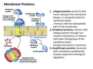
Membrane proteins normal the reduced LD, LDr = LD/isotropic absorbance, is: where is the angle the transition moment of interest makes with the normal to the cylinder surface (i.e. to the lipid long axis), S is the orientation factor that denotes the fraction of the liposome that is oriented as a cylinder perfectly parallel to the flow direction.
-helix 220 nm n-* polarised helix axis 208 nm // helix axis Helix // lipids at 208 nm LD<0, 220 nm >0 Gramicidin
LD of nanotubes • Single walled nanotubes (as received) • UV spectroscopy: ? p-plasmon band Abs of SWNT In SDS < CMC LD of SWNT + anthracene LD of anthracene expanded LD of SWNT
DNA binding modes 1. Intercalation between base pairs: DNA lengthens and stiffens 2. In major groove: more options for selectivity 3. In minor groove: planar aromatics with AT rich DNA 4. External binding: non specific
DNA binding & structure control by transition metal supramolecular helicates Alison Rodger and Mike Hannon Isabelle Meistermann, Karen Sanders, Chris Isaac, Jemma Peberdy, Laura Childs, Syma Khalid, Mark Rodger University of Warwick Virtudes Moreno, Barcelona (AFM) Einar Sletten, Norway (NMR)
An inexpensive approach Simple imines can be used for supramolecular assembly e.g. FeCl2 • Yield 87% • Isolate by filtration • Cost per gramme: 8p (0.8FFr; • 0.13 US$; 0.23DM; 230ItLi; 50GrD) X-ray structure M.J. Hannon, C.L. Painting, A. Jackson, J. Hamblin, and W. Errington, Chem. Commun., 1997, 1807.
Probing binding by absorbance • Only very small changes observed in the MLCT bands at ~ 580 nm. • Larger changes in ligand based band at ~ 330nm. • Changes confirm binding. • Small changes suggest binding does not cause major perturbations in the helical structure. Absorbance spectra of ct-DNA with 20 mM [Fe2L3]Cl4
Stretched film LD of the di-iron helicate • MLCT bands at ~580 nm are radially (xy) polarised. • The ligand band at the ~320 nm contains both radial (xy) and long axis (z) contributions • Aim to understand flow LD … LD of spectra of [Fe2L3]Cl4 in a stretched polyvinylalcohol film
Kinking the DNA The flow LD experiment not only reveals a binding angle of 60°(±10 °) but also reveals changes in the DNA region. The decrease in the magnitude of the LD signal in the DNA region with respect to free DNA means a decrease in DNA orientation. This indicates that the [Fe2L3]Cl4 helicate is kinking the DNA. LD spectra of free and bound ct-DNA
AFM studies with linear DNA with complex no complex mm mm And more more mm mm
free DNA DNA:M = 10:3 DNA:P = 10:3 DNA:rac = 10:3 AFM images of a fragment of 200 pb linear DNA incubated at 37ºC for 5 h with the iron cylinders.