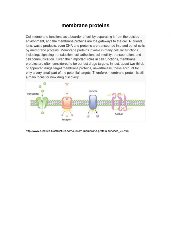
Membrane Proteins
Membrane Proteins. Fig. 10-10 in FOB. Membrane Proteins. Classes (kind of membrane association): Transmembrane : rule: polypeptide chain passes completely through the bilayer examples: single membrane-spanning domain: hydrophobic a -helix (e.g., glycophorin)

Membrane Proteins
E N D
Presentation Transcript
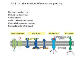
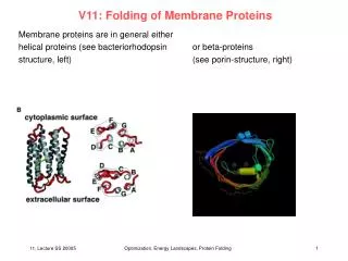
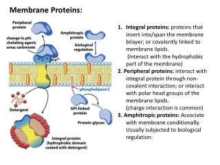
Membrane Proteins • Classes (kind of membrane association): • Transmembrane: • rule: polypeptide chain passes completely through the bilayer • examples: • single membrane-spanning domain: hydrophobic a-helix (e.g., glycophorin) • multiple membrane-spanning domains (e.g., 7-pass transmembrane proteins such as bacteriorhodopsin) • hydropathy plots to detect transmembrane domains • pores: a-helical vs. b-sheet/barrel (porins)
1. Proteins that span a membrane have a characteristic structure in the region of the lipid bilayer. Which, if any, of the three 20-amino acid sequences listed below is the most likely candidate for such a transmembrane segment? Explain the reasons for your choice.A. Ile Thr Leu Ile Tyr Phe Gly Val Met Ala Gly Val Ile Gly Thr Ile Leu Leu Ile Ser B. Ile Thr Pro Ile Tyr Phe Gly Pro Met Ala Gly Val Ile Gly Thr Pro Leu Leu Ile Ser C. Ile Thr Glu Ile Tyr Phe Gly Arg Met Ala Gly Val Ile Gly Thr Asp Leu Leu Ile Ser
Membrane Proteins • Classes (kind of membrane association): • Lipid anchoring (covalent attachment to membrane phospholipid): • For proteins on cytosolic face: • fatty acid: • myristate (C14): amide linkage to amino group of N-terminal glycine • palmitate (C16): thioester linkage to Cys residue • prenylation: e.g. polyisoprenoid (farnesyl or geranylgeranyl group) linked to methylated C-terminal Cys (which was initially four residues from C-terminus, but last three residues are cleaved off, then carboxyl group of Cys is methylated) • For proteins on exoplasmic face: • GPI (glycosylphosphatidylinositol) anchor: amide linkage to C-terminal residue of protein • Peripheral: non-covalent protein-protein interactions w/ integral membrane protein(s). The bulk of the peripheral membrane protein resides entirely on one face of the membrane
Many membrane proteins display rotational and lateral diffusion, but they are much slower than phospholipids (1/10 - 1/100 the rate) • Experimental evidence for rapid redistribution of membrane proteins: • Making heterokaryons – e.g., fusing mouse and human cells; specific cell surface antigens unique to either mouse or human cells were recognized by differentially-labeled antibodies. With time, mixing of cell surface antigens was observed. • Patching and capping – multivalent ligands that recognize specific membrane proteins bind to and cross-link the proteins, which aggregate into clusters (patching); patches are then actively swept to one end of the cell (capping). • Fluorescence recovery after photobleaching (FRAP) – membrane proteins are labeled with a fluorescent molecule; illumination of a small area of the cell surface bleaches out the fluorescence; over time, fluorescent molecules from adjoining unbleached areas are seen to move into the bleached area. Can be used to determine rates of lateral diffusion.
Factors that limit the diffusion of membrane proteins: • Attachment to the cytoskeleton and/or extracellular matrix. • Assembly of cell-cell adhesion complexes and other cell junctions. • Tight junctions isolate basal-lateral surfaces from apical surfaces in polarized epithelia.
Membrane Carbohydrates • Carbohydrates especially abundant on plasma membrane; glycocalyx formed of glycolipids, glycoproteins and proteoglycans. • Sugars can be attached to proteins (almost all) or lipids (1 out of 10). • Asymmetric: only extracellular face of plasma membrane is glycosylated. • Functions: • Protection: important for lubrication and structural integrity of the cell surface. • Important for certain cell-cell recognition events (ex. WBC adhesion to endothelial lining of blood vessels).