Download
1 / 24
240 likes | 265 Views
Understand the levels of protein structure, from primary to quaternary, and their role in determining protein function. Learn about the forces that stabilize protein structure and the techniques used to determine protein conformation. Delve into secondary structures like α-helix and β-pleated sheet, and discover the diversity of protein shapes, including fibrous and globular proteins.

E N D
Polypeptide and protein structure Nafith Abu Tarboush DDS, MSc, PhD natarboush@ju.edu.jo www.facebook.com/natarboush
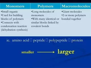
Protein conformation • Many conformations are possible for proteins due to flexibility of amino acids linked by peptide bonds • At least one major conformations has biological activity, and hence is considered the protein’s native conformation or the native protein
Levels of Protein Structure • 1°structure: sequence and number, from N to C • 2°structure: the ordered 3-dimensional arrangements (conformations) in localized regions of a polypeptide chain, backbone interactions through hydrogen bonding; • e. g., -helix and -pleated sheet • 3˚ structure: 3-D arrangement of all atoms • 4˚ structure: arrangement of monomer subunits with respect to each other
Primary Structure of Proteins • Zigzag arrangement • R-groups • Determination? • 1o Sequence & 3-D conformation; relation to functional properties (MW& genetic mutations) • Site-directed mutagenesis and structure function relationship
Shape-Determining Interactions in Proteins • Is it ordered or spaghetti? • Hydrogen, ionic, covalent, & hydrophobic
Secondary Structure of Proteins • What is the secondary structure of proteins? “folding of the backbone” • What are the bonds that have a free rotation? What is the implication? These angles repeat themselves in regular secondary structures • Two main kinds: α-helix and β-pleated sheet • They are periodic; their features repeat at regular intervals • Stability of secondary structure
The α-helix • H-bonds are parallel to the helix axis, same segment • C=O binds N—H four residues away • linear arrangement of H-bonds (maximum strength and stability) • Turns occur every 3.6 residues, right handed, clockwise • The pitch (linear distance between corresponding points on successive turns) is 5.4 Å • Proteins have varying amounts of α-helical structures • What factors affect the helix (specific amino acids, electrostatic repulsion, steric repulsion)
The β-sheets • Backbone is almost completely extended • R groups extending above and below the sheet • H-bonds are intra-chain or inter-chain bonds • Perpendicular to the direction of the protein chain • Parallel vs. anti-parallel • Zigzag structure
Others regular ones • Why do we have them? • Loops vs. turns • Also known as β-bends • Involve 4 amino acids (H-bond: C=O of 1 & N–H of 4) • A motif is a repetitive super-secondary structure. Regardless of function • Domains; protein conformations with similar functions (leucine zipper)
-Helices and -Sheets • Supersecondary structures: a combination of - and -sections • b unit: (parallel) • unit: (helix-turn-helix), anti-parallel • -meander: an anti-parallel sheet formed by a series of tight reverse turns connecting stretches of a polypeptide chain • Greek key: a repetitive super-secondary structure formed when an anti-parallel sheet doubles back on itself • -barrel: created when -sheets are extensive enough to fold back on themselves
Fibroin, β-sheets, alternating glycine and alanine α-keratins, bundles of α-helices Fibrous Proteins • Contain polypeptide chains organized approximately parallel along a single axis: • Consist of long fibers or large sheets • Mechanically strong • Insoluble • play an important structural role • Examples are • Keratin • Collagen • fibroin
Globular Proteins • Folded to, a more or less, spherical shape • Soluble • Polar vs. non-polar, exterior vs. interior • Most of them have substantial sections of -helix and -sheet
3˚ Structure • The 3-dimensional arrangement of all the atoms in the molecule • Simple vs. conjugated • Backbone hydrogen bonding, side chains hydrogen bonding, hydrophobic interactions, electrostatic attraction, electrostatic repulsion, metal coordination • Not every protein have all kinds of interactions (myoglobin & hemoglobin; no disulfides) (trypsin & chymotrypsin, no metal complexes) • Interactions between side chains also plays a role
3°& 4°Structure • Tertiary (3°) structure: the arrangement in space of all atoms in a polypeptide chain • It is not always possible to draw a clear distinction between 2°and 3°structure • Quaternary (4°) structure: the association of polypeptide chains into aggregations (dimers, trimers, tetramers,…etc). Non-covalent interactions. Simple or conjugated
Determination of 3°Structure • X-ray crystallography • uses a perfect crystal; that is, one in which all individual protein molecules have the same 3D structure and orientation • exposure to a beam of x-rays gives a series diffraction patterns • information on molecular coordinates is extracted by a mathematical analysis called a Fourier series • 2-D Nuclear magnetic resonance • can be done on protein samples in aqueous solution
X-Ray and NMR Data • High resolution method to determine 3˚ structure of proteins (from crystal) • Diffraction pattern produced by electrons scattering X-rays • Series of patterns taken at different angles gives structural information • Determines solution structure • Structural info. Gained from determining distances between nuclei that aid in structure determination • Results are independent of X-ray crystallography
Complex Protein Structures • Covalent conjugation to carbohydrates (glyco-proteins): post-translational, function, N-linked (-N of Asn) & O-linked (-OH of Ser or Thr) & occasionally to the –OH of hydroxy-lysine • Lipoproteins: Non-covalent, store & transport lipids and cholesterol • Extremely important: ex. Erythrocytes & blood groups (glycoshpingolipids) • Phosphoproteins: contain phosphate groups esterified to Ser, Thr, or Tyr.; the phosphate group usually regulates protein function
Chemical Properties of Proteins • 1. Protein Hydrolysis • The reverse of protein synthesis • Digestion of proteins is hydrolyzing peptide bonds • Takes place in the stomach and small intestine
2. Protein Denaturation • How the protein preserve its shape? • What is denaturation? It affects physical, chemical, and biological properties, such as enzymes • Solubility decreased, ex. eggs • Heat (≈≥50 οC) • Mechanical agitation • Detergents • Organic compounds: acetone, ethanol, bacterial proteins • pH change: disrupt salt bridges, milk turns sour because it has become acidic • Most denaturation is irreversible
Protein Folding & prediction • The integration of biochemistry and computing has led to bioinformatics • Predicting the 3˚ structure (limitations) • First step is to search for sequence homology
Hydrophobic Interactions & Chaperons • Hydrophobic interactions are spontaneous • Chaperones aid in correct & timely folding of many proteins • hsp70 were the first chaperone proteins discovered • Chaperones exist in organisms from prokaryotes to humans
Structure & diseases (prion, Alzheimer) • Mad-cow disease (bovine spongiform encephalopathy), and human spongiform encephalopathy (Creutzfeldt-Jakob disease) • The cause is a small (28-kDa) protein called a prion (Met129) • Alzheimer's Disease: buildups of plaques and fibers of hard insoluble material • Amyloid protein mis-folded and broken down (≈40 a.a, Ab protein) and aggregates