Download
1 / 22
220 likes | 246 Views
Explore the fabrication and purpose of 3D microfluidic devices for studying chemotaxis in metastatic breast cancer cells, aiming to improve diagnosis and treatment. Learn about the fabrication steps, challenges, and successful experiments achieved. Thank you to all contributors!

E N D
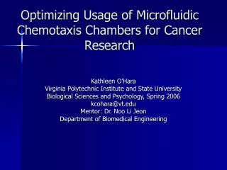
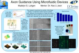
Fabrication of Microfluidic Devices for 3D Chemotaxis Studies Delaram Sahebzamani University of South Florida Mentor: Noo Li Jeon Department of Biomedical Engineering
Jeon Lab The lab’s focus is on applying microfluidics to biology. For the past few years they have concentrated on studying chemotaxis of metastatic breast cancer cells.
Chemotaxis • In chemotaxis cells exhibit directed movement in response to external concentration gradients. • By studying the migration of these breast cancer cells in precisely controlled gradients, one can gain further insights into processes that will eventually lead to better diagnosis and treatment.
The Next Step • All experiments done to this point have been 2 dimensional and the NEXT STEP is to develop devices to study migration 3D.
The Answer • In various incidents cells migrate 3 dimensionally and it is important to create such environments. • The tissues and organs of the human body are abundant with Collagen. Therefore by using Collagen Type 1 within the microfluidic devices will allow us to mimic in vivo cells. • In vitro studies should mimic in vivo environment.
Fabrication Steps • Plasma the Device onto a Glass Slide • Inject Collagen solution into device through inlet • Suction from outlet and allow for polymerization to occur
Problems • The Texture • Pressure Flow
L W H Cell Density Experiments • 2 Cell Lines: MDA MB 231 & MTLn3 • 30,000 • 40,000 • 70,000 • 100,000
100,000 MDA MB 231 Cells Phase 10X
40,000 MTLn3 Cells Phase 10X Fluorescence 10X
70,000 MTLn3 Cells Phase 10X Fluorescence10x
30,000 MTLn3 with Confocal Microscope 300 Microns, Every 10 Microns
Summary of Cell Density • Middle channels are 300 microns in depth and it was determined that 30,000 to 40,000 cells was approximately the right range to use for future experiments
Conclusion • 3D Microfluidic Device was in fact developed!!!!!!!!! • These new 3D devices will now be used for chemotaxis studies.
Acknowledgements • Mentor: Dr. Noo Li Jeon • Graduate Student: Wajeeh Saadi • Lab: Jeong Won Park , Anne Taylor, Bonggeun Chung, Kathleen O’ Hara, Babak Mosadegh, Madelyn Luttgen, Cyrus Roushan • IM SURE: Said Shokair, Jerry McMillan, Goran Matijasevic • National Science Foundation