Download
1 / 26
310 likes | 578 Views
Diffusion model fitting and tractography: A primer. Anastasia Yendiki HMS/MGH/MIT Athinoula A. Martinos Center for Biomedical Imaging. White-matter imaging. Axons measure ~ m in width They group together in bundles that traverse the white matter

E N D
Diffusion model fitting and tractography: A primer Anastasia Yendiki HMS/MGH/MIT Athinoula A. Martinos Center for Biomedical Imaging Why’n’how | Diffusion model fitting and tractography
White-matter imaging • Axons measure ~m in width • They group together in bundles that traverse the white matter • We cannot image individual axons but we can image bundles with diffusion MRI • Useful in studying neurodegenerative diseases, stroke, aging, development… From the National Institute on Aging From Gray's Anatomy: IX. Neurology Why’n’how | Diffusion model fitting and tractography
Diffusion in brain tissue • Differentiate tissues based on the diffusion (random motion) of water molecules within them • Gray matter: Diffusion is unrestricted isotropic • White matter: Diffusion is restricted anisotropic Why’n’how | Diffusion model fitting and tractography
How to describe diffusion • At every voxel we want to know: • Is this in white matter? • If yes, what pathway(s) is it part of? • What is the orientation of diffusion? • What is the magnitude of diffusion? • A grayscale image cannot capture all this! Why’n’how | Diffusion model fitting and tractography
Diffusion MRI Diffusion encoding in direction g1 • Magnetic resonance imaging can provide “diffusion encoding” • Magnetic field strength is varied by gradients in different directions • Image intensity is attenuated depending on water diffusion in each direction • Compare with baseline images to infer on diffusion process g2 g3 g4 g5 g6 No diffusion encoding Why’n’how | Diffusion model fitting and tractography
Need to know: Gradient directions • True diffusion direction || Applied gradient direction Maximum attenuation • True diffusion direction Applied gradient direction No attenuation • To capture all diffusion directions well, gradient directions should cover 3D space uniformly Diffusion-encoding gradient g Diffusion detected Diffusion-encoding gradient g Diffusion not detected Diffusion-encoding gradient g Diffusion partly detected Why’n’how | Diffusion model fitting and tractography
How many directions? • Acquiring data with more gradient directions leads to: • More reliable estimation of diffusion measures • Increased imaging time Subject discomfort, more susceptible to artifacts due to motion, respiration, etc. • DTI: • Six directions is the minimum • Usually a few 10’s of directions • Diminishing returns after a certain number [Jones, 2004] • HARDI/DSI: • Usually a few 100’s of directions Why’n’how | Diffusion model fitting and tractography
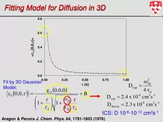
Need to know: b-value • The b-value depends on acquisition parameters: b = 2G22 (- /3) • the gyromagnetic ratio • G the strength of the diffusion-encoding gradient • the duration of each diffusion-encoding pulse • the interval b/w diffusion-encoding pulses 90 180 acquisition G Why’n’how | Diffusion model fitting and tractography
How high b-value? • Increasing the b-value leads to: • Increased contrast b/w areas of higher and lower diffusivity in principle • Decreased signal-to-noise ratio Less reliable estimation of diffusion measures in practice • DTI: b ~ 1000 sec/mm2 • HARDI/DSI: b ~ 10,000 sec/mm2 • Data can be acquired at multiple b-values for trade-off • Repeat acquisition and average to increase signal-to-noise ratio Why’n’how | Diffusion model fitting and tractography
Looking at the data A diffusion data set consists of: • A set of non-diffusion-weighted a.k.a “baseline” a.k.a. “low-b” images (b-value = 0) • A set of diffusion-weighted (DW) images acquired with different gradient directions g1, g2, … and b-value >0 • The diffusion-weighted images have lower intensity values b2, g2 b3, g3 b=0 b1, g1 Baseline image Diffusion-weighted images b4, g4 b5, g5 b6, g6 Why’n’how | Diffusion model fitting and tractography
Data analysis steps • Pre-process images • FSL: eddy_correct, rotate_bvecs • Fit a diffusion model at every voxel • DTK: DSI, Q-ball, or DTI • FSL: Ball-and-stick (bedpost) or DTI (dtifit) • Compute measures of anisotropy/diffusivity and compare them between populations • Voxel-based, ROI-based, or tract-based statistical analysis • For tract-based: Reconstruct pathways • DTK: Deterministic tractography using DSI, Q-ball, or DTI model • FSL: Probabilistic tractography (probtrack) using ball-and-stick model DTK: www.trackvis.org, FSL: www.fmrib.ox.ac.uk/fsl Why’n’how | Diffusion model fitting and tractography
Models of diffusion Why’n’how | Diffusion model fitting and tractography
A bit more about the tensor • A tensor can be thought of as an ellipsoid • It can be defined fully by: • 3 eigenvectorse1, e2, e3 (orientations of ellipsoid axes) • 3 eigenvalues1 , 2, 3 (lengths of ellipsoid axes) 1e1 2e2 3e3 Why’n’how | Diffusion model fitting and tractography
Tensor: Physical interpretation • Eigenvectors express diffusion direction • Eigenvalues express diffusion magnitude Isotropic diffusion: 123 • Anisotropic diffusion: • 1>>2 3 1e1 1e1 2e2 3e3 2e2 3e3 Why’n’how | Diffusion model fitting and tractography
Tensor: Summary measures • Mean diffusivity (MD): • Mean of the 3 eigenvalues Faster diffusion Slower diffusion MD(j) = [1(j)+2(j)+3(j)]/3 • Fractional anisotropy (FA):Variance of the 3 eigenvalues, normalized so that0 (FA) 1 Anisotropic diffusion Isotropic diffusion [1(j)-MD(j)]2+ [2(j)-MD(j)]2+ [3(j)-MD(j)]2 3 FA(j)2 = 1(j)2+ 2(j)2+ 3(j)2 2 Why’n’how | Diffusion model fitting and tractography
Tensor: More summary measures • Axial diffusivity: Greatest eigenvalue • Radial diffusivity: Average of 2 lesser eigenvalues • Inter-voxel coherence: Average angle b/w the major eigenvector at some voxel and the major eigenvector at the voxels around it AD(j) = 1(j) RD(j) = [2(j) + 3(j)]/2 Why’n’how | Diffusion model fitting and tractography
Tensor: Visualization Image: An intensity valueat each voxel Tensor map: A tensorat each voxel Direction of eigenvector corresponding to greatest eigenvalue Why’n’how | Diffusion model fitting and tractography
Tensor: Visualization Image: An intensity valueat each voxel Tensor map: A tensorat each voxel Direction of eigenvector corresponding to greatest eigenvalue Red: L-R, Green: A-P, Blue: I-S Why’n’how | Diffusion model fitting and tractography
? Tractography • Use local diffusion orientation at each voxel to determine pathway between distant brain regions • Local orientation comes from diffusion model fit (tensor, ball-and-stick, etc.) • Deterministic vs. probabilistic tractography: • Deterministic assumes a single orientation at each voxel • Probabilistic assumes a distribution of orientations • Local vs. global tractography: • Local fits the pathway to the data one step at a time • Global fits the entire pathway at once Why’n’how | Diffusion model fitting and tractography
Deterministic vs. probabilistic • Deterministic methods give you an estimate of model parameters 5 • Probabilistic methods give you the uncertainty (probability distribution) of the estimate 5 Why’n’how | Diffusion model fitting and tractography
Deterministic vs. probabilistic Sample 1 … Sample 2 Deterministic tractography: One streamline per seed voxel Probabilistic tractography: Multiple streamline samples per seed voxel (drawn from probability distribution) Why’n’how | Diffusion model fitting and tractography
Deterministic vs. probabilistic Deterministic tractography: One streamline per seed voxel Probabilistic tractography: A probability distribution (sum of all streamline samples from all seed voxels) Why’n’how | Diffusion model fitting and tractography
Local vs. global Local tractography: Fits pathway step-by-step, using local diffusion orientation at each step Global tractography: Fits the entire pathway, using diffusion orientation at all voxels along pathway length Why’n’how | Diffusion model fitting and tractography
Local tractography • Best suited for exploratory study of connections • All connections from a seed region, not constrained to a specific target region • How do we isolate a specific white-matter pathway? • Thresholding? • Intermediate masks? • Non-dominant connections are hard to reconstruct • Results are not symmetric between “seed” and “target” regions • Sensitive to areas of high local uncertainty in orientation (e.g., pathaway crossings), errors propagate from those areas Why’n’how | Diffusion model fitting and tractography
Global tractography • Best suited for reconstruction of known white-matter pathways • Constrained to connection of two specific end regions • Not sensitive to areas of high local uncertainty in orientation, integrates over entire pathway • Symmetric between “seed” and “target” regions • Need to search through a large solution space of all possible connections between two regions: • Computationally expensive • Sensitive to initialization Why’n’how | Diffusion model fitting and tractography
TRACULA • TRActs Constrained by UnderLying Anatomy • Automatic reconstruction of probabilistic distributions of 18 major white-matter pathways • No manual labeling of ROIs needed • Use prior information on pathway anatomy from training data: • Manually labeled pathways in training subjects • FreeSurfer segmentations of same subjects • Learn neighboring anatomical labels along pathway • Beta version available in FreeSurfer 5.1 Why’n’how | Diffusion model fitting and tractography