Protein Structure
510 likes | 760 Views
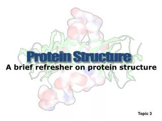
Protein Structure. A brief refresher on protein structure. Topic 3. Perhaps the most important structural bioinformatics result ever published…. Chothia , C. & Lesk , A. M. (1986). The relation between the divergence of sequence and structure in proteins. E MBO J. , 5(4):823-826. .

Protein Structure
E N D
Presentation Transcript
Protein Structure A brief refresher on protein structure Topic 3
Perhaps the most important structural bioinformatics result ever published… Chothia, C. & Lesk, A. M. (1986). The relation between the divergence of sequence and structure in proteins.EMBO J., 5(4):823-826. Starting now, the relationships identified by this simple graph will impact everything we do throughout the remainder of the class.
Levels of Protein Structures • primary structure • (set of covalent bonds within the structure) • secondary structure (helices, strands, coils/loops) • tertiary structure (3D packing of secondary structures) • quaternary structure • (spatial arrangements of multiple chains) LIRLFKSHPETLEKFDRFKHL…
The three most common classes of proteins Globular protein Fibrous protein Membrane protein
Formation of a Peptide Bond by Condensation Amino Acid 1 Amino Acid 2 Note: this chemistry will not work as drawn! Peptide bond Peptide bond is the amide linkage that is formed between two amino acids, which results in (net) release of a molecule of water (H2O). The four atoms in the yellow box form a rigid planar unit and, as we will see next, there is no rotation around the C-N bond.
The primary structure is the set of all covalent bonds within the protein, which is approximated by the sequence (-CSS). Note: Primary structure or sequence, but not primary sequence.
Peptides Q: why is the pentapeptide SGYAL different than LAYGS?
A Closer Look at the Peptide Bond • The carbonyl group has a partial negative charge and the amide nitrogen has a partial positive charge, which set up a small electric dipole. • The peptide C-N bond has a partial double-bond character (partial sharing of two pairs of electrons between O and N. • The peptide bond is planar and rigid, cannot rotate freely (see next slide). • Resonance: delocalization of bonding electrons over more than one chemical bond. Lehninger Principles of Biochemistry
A Closer Look at the Peptide Bond • But, bonds N-C and C -C can rotate, which can be described by two torsion angles: (phi) and (psi). • (phi): C-N-C-C • (psi): N-C-C-N • (omega): C-C-N- C • In principle, and can have any value between -180O and +180O. But due to steric interference……
A Closer Look at the Peptide Bond Bond lengths : C -C C-N N-C The peptide bond is normally in the trans configuration (~99.6% of the time) = 180O One exception is for proline. The fraction of X-Pro peptide bonds in the cis isomer under unstrained conditions ranges from 10-40%. The fraction depends slightly on the preceding amino acid X. Note the differences between C-N and N-C
An (a.) unstable vs. (b.) the most stable Ala-Ala dipeptide conformation (a.) (b.)
The rotatable backbone torsion angles Rotation of phi Rotation of psi
The Ramachandran plot With a molecular modeling kit, prove to yourself that (0,0) is an unallowable due to a steric clash.
Prolylcis-transisomerization as a molecular switch “The local environment of proline within a protein can influence the relative free energies of the cis and trans isomeric states, leading to wide variations in the ratio of cis:trans populations in different proteins. Although most structures require proline to adopt one or the other isomer in the context of native protein folds, several recent structures show the presence of both populations for specific proline residues.” Liu et al. Nature Chemical Biology. 3, 619 - 629 (2007)
Final thoughts on primary structure -- The primary structure is a complete description of the covalent bond network within a protein. -- This is almost(!) completely described by the sequence of amino acids. -- If you know that the protein is AVG…, you can look up the structures of A, V and G, plus what you know about peptide bonding allows you to complete the covalent bond structure. -- So, when does the primary structure not fully describe the covalent bond network?
Secondary structure = local regions of proteins characterized by (i.) similar f/y values and (ii.) backbone hydrogen bonding
Hydrogen Bond H-bond donor H-bond acceptor • A weak bond involving the sharing of an electron with a hydrogen atom Common hydrogen bonds in biological systems Directionality of the H-bond
Secondary Structure:-helix 5.4 Å 3.6 residue = -57O, = -47O Hydrogen bond pattern: C=O (i) and N-H (i+4) Image from “Protein Structure and Function” by Gregory A Petsko and Dagmar Ringe
Protein Secondary Structure: helices • Other helical conformations • 310 helix, • helix =-49O, =-26O Hydrogen bond pattern: C=O (i) and N-H (i+3) Residues per turn: 3 =-57O, =-70O Hydrogen bond pattern: C=O (i) and N-H (i+5) Residue per turn: 4.4
Amphipathichelix hydrophobic Hydrophilic protein packing and function Image from “Protein Structure and Function” by Gregory A Petsko and Dagmar Ringe
-sheet (Pleated Sheet) Parallel -sheet Anti-parallel -sheet 6.5Å 7Å = -119O, = +113O = -139O, = +135O Lehninger Principles of Biochemistry
Amino acid propensity Used in the first generation of secondary structure prediction methods, e.g. Chou-Fasman
-turns • the distance between the C atom of residue i and the C atom of residue i+3 • is less than 7Å • the central two residues are not helical • on the basis of the phi, psi angles of residues i+1 and i+2
-turns Turn propensities (Ft/Fb) Lehninger Principles of Biochemistry
Ramachandran Plot Red: allowed regions Yellow: additionally allowed regions White: disallowed regions
Not all Ramachandran plots are created equal. THE 18 STANDARD AMINO ACIDS GLYCINE PROLINE
Tertiary structure = the 3D shape of a single protein chain, which is stabilized by a large number of noncovalent interactions.
Ruminations on protein stability... • Protein stability is a small difference of large numbers. • Proteins are stable (G < 0) only over a narrow environmental range. • In fact, there are forces pushing the equilibrium between folded and unfolded in both directions. • Stabilizing forces: Intraprotein salt bridges, hydrogen bonds, dipole-dipole interactions and VDW interactions (all of which are electrostatic in nature). • Destabilizing forces: Primarily electrostatic interactions with solvent and conformational entropy reduction.
There are many different ways to represent a protein structure
It is common, and sometimes useful, to think of protein structures as scaffolding upon which ‘active sites’ are attached.
Conformational changes within the tetramer structure underlie the differences in oxygen affinity between oxy- and deoxy-hemoglobin.


