Download
1 / 14
170 likes | 462 Views
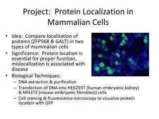
Project: Protein Localization in Mammalian Cells. Idea: Compare localization of proteins (ZFP568 & GALT) in two types of mammalian cells Significance: Protein location is essential for proper function, mislocalization is associated with disease. Biological Techniques:

E N D
Project: Protein Localization in Mammalian Cells • Idea: Compare localization of proteins (ZFP568 & GALT) in two types of mammalian cells • Significance: Protein location is essential for proper function, mislocalization is associated with disease • Biological Techniques: • DNA extraction & purification • Transfection of DNA into HEK293T (human embryonic kidney) & NIH3T3 (mouse embryonic fibroblast) cells • Cell staining & fluorescence microscopy to visualize protein location with GFP
Diseases associated with defects in protein transport • Cystic fibrosis (CF) • Familial hypercholesterolaemia (FH) • Congenital sucrase-isomaltase deficiency (CSID)
Project Goal • Compare the localization of two proteins (ZFP568 & GALT) in two types of mammalian cells (human embryonic kidney cells & mouse embryonic fibroblasts) • We’ll do this by • Making DNA that codes for our proteins (DNA extraction & purification) • Putting that DNA in mammalian cells (transfection) • Determining where our proteins are within the cells (cell staining & fluorescent microscopy)
Our Project Plan Stain & look at our cells to see where the protein is (Thurs/Fri) Miniprep to extract DNA from bacteria (Tues) Transfect to put our DNA in mammalian cells (Wed)
The Two Cell Types We’ll Use • HEK293T cells (human embryonic kidney) • NIH 3T3 cells (mouse embryonic fibroblast) • Look at the cell morphology of each: How are they different? • Which cell type wouldyou want to use for our project?
The First Protein We’ll Look At • GFP-ZFP568 (Zinc finger protein 568) • Binds to DNA and recruits transcriptional repressor TRIM28 • Mutation (“chato”) causes embryonic arrest • Garcia-Garcia, M.J., Shibata, M., and Anderson, K.V. (2008). Development135, 3053-3062.
The Second Protein We’ll Look At • GFP-GALT (Galactose-1-phosphate uridylyltransferase) • Enzyme important in sugar metabolism: Converts galactose to glucose • Mutation in GALT causes galactosemia • autosomal recessive mode of inheritance
GFP vector – a plasmid DNA coding for ZFP568 or GALT can be inserted here
Restriction Enzymes • XhoI and HindIII • Used to cut (digest) DNA coding for ZFP568 so it could be glued (ligated) into GFP-vector • Digest with these enzymes and DNA coding for ZFP568 should be “cut” from its vector • Resulting fragments of DNA will be analyzed using gel electrophoresis • What will this gel look like? How will digested GFP-ZFP568 look different from GFP-alone? • XhoI • HindIII
DNA extraction & purification • GFP-ZFP568, GFP-GALT, and GFP-alone DNA was transformed into bacterial cells cells were cultured to make more DNA now DNA can be extracted • QiagenMiniPrep Kit
Transfection • Purified DNA can be transfected into HEK293T and NIH3T3 cells using Lipofectamine 2000 • Mix DNA in media with the Lipofectamine reagent and then add it to your cells’ dish • Cells will take up DNA and express the proteins (GFP-ZFP568, GFP-GALT, or GFP-alone) within 24hrs • Why do we transfect the GFP-alone construct?
Cell Staining • Phalloidin (red) • Marks actin filaments, concentrated beneath cell membrane to keep cell shape • Actin is part of cytoskeleton • DAPI (blue) • Marks the nuclei of cells • GFP (green) • Tagged to ZFP568 and GALT • Can take pictures of each using fluorescence microscope and then merge using Photoshop • Why do we stain with DAPI and Phalloidin? • Why don’t we have to stain to see ZFP568 or GALT? http://migration.wordpress.com/2007/07/11/basics-the-cytoskeleton/
Final Questions • Where is GFP-GALT and GFP-ZFP568 located within the cell? Why? • What does GFP-alone look like? Why? • Is protein location different in HEK293T or NIH3T3 cells? • Why are cells useful for scientists?