Download

1 / 103
1.11k likes | 1.79k Views
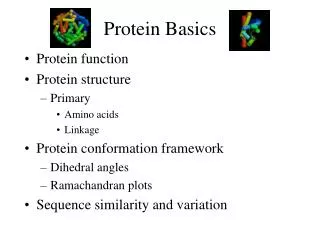
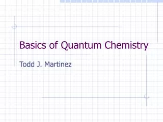
Protein Chemistry Basics. Protein function Protein structure Primary Amino acids Linkage Protein conformation framework Dihedral angles Ramachandran plots Sequence similarity and variation. Protein Function in Cell. Enzymes Catalyze biological reactions Structural role Cell wall

E N D
Protein ChemistryBasics • Protein function • Protein structure • Primary • Amino acids • Linkage • Protein conformation framework • Dihedral angles • Ramachandran plots • Sequence similarity and variation
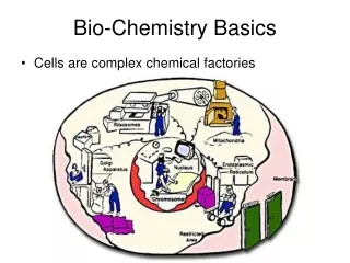
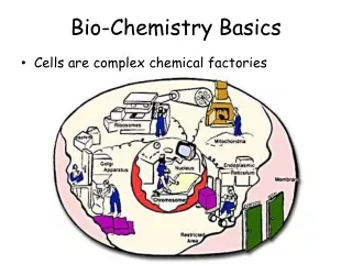
Protein Function in Cell • Enzymes • Catalyze biological reactions • Structural role • Cell wall • Cell membrane • Cytoplasm
Hemoglobin: Background • Protein in red blood cells
Hemoglobin: Background • Protein in red blood cells • Composed of four subunits, each containing a heme group: a ring-like structure with a central iron atom that binds oxygen
Hemoglobin: Background • Protein in red blood cells • Composed of four subunits, each containing a heme group: a ring-like structure with a central iron atom that binds oxygen • Picks up oxygen in lungs, releases it in peripheral tissues (e.g. muscles)
Hemoglobin – Quaternary Structure Two alpha subunits and two beta subunits (141 AA per alpha, 146 AA per beta)
Hemoglobin – Tertiary Structure One beta subunit (8 alpha helices)
Hemoglobin – Secondary Structure alpha helix
β-Hairpin Motif • Simplest protein motif involving two beta strands [from Wikipedia] • adjacent in primary sequence • antiparallel • linked by a short loop • As isolated ribbon or part of betasheet • a special case of a turn • direction of protein backbone reverses • flanking secondary structure elements interact (hydrogen bonds) CS 882 course project
Types of Turns • β-turn (most common) • donor and acceptor residues of hydrogen bonds are separated by 3 residues (ii +3 H-bonding) • δ-turn • ii +1 H-bonding • γ-turn • ii +2 H-bonding • α-turn • ii +4 H-bonding • π-turn • ii +5 H-bonding • ω-loop • a longer loop with no internal hydrogen bonding CS 882 course project
Structure Stabilizing Interactions • Noncovalent • Van der Waals forces (transient, weak electrical attraction of one atom for another) • Hydrophobic (clustering of nonpolar groups) • Hydrogen bonding
Hydrogen Bonding • Involves three atoms: • Donor electronegative atom (D) (Nitrogen or Oxygen in proteins) • Hydrogen bound to donor (H) • Acceptor electronegative atom (A) in close proximity D – H A
δ- δ+ δ- D – H A D-H Interaction • Polarization due to electron withdrawal from the hydrogen to D giving D partial negative charge and the H a partial positive charge • Proximity of the Acceptor A causes further charge separation
δ- δ+ δ- D – H A D-H Interaction • Polarization due to electron withdrawal from the hydrogen to D giving D partial negative charge and the H a partial positive charge • Proximity of the Acceptor A causes further charge separation • Result: • Closer approach of A to H • Higher interaction energy than a simple van der Waals interaction
Hydrogen Bonding And Secondary Structure beta-sheet alpha-helix
Structure Stabilizing Interactions • Noncovalent • Van der Waals forces (transient, weak electrical attraction of one atom for another) • Hydrophobic (clustering of nonpolar groups) • Hydrogen bonding • Covalent • Disulfide bonds
Disulfide Bonds • Side chain of cysteine contains highly reactive thiol group • Two thiol groups form a disulfide bond
Disulfide Bonds • Side chain of cysteine contains highly reactive thiol group • Two thiol groups form a disulfide bond • Contribute to the stability of the folded state by linking distant parts of the polypeptide chain
Hemoglobin – Primary Structure NH2-Val-His-Leu-Thr-Pro-Glu-Glu- Lys-Ser-Ala-Val-Thr-Ala-Leu-Trp- Gly-Lys-Val-Asn-Val-Asp-Glu-Val- Gly-Gly-Glu-….. beta subunit amino acid sequence
Protein Structure - Primary • Protein: chain of amino acids joined by peptide bonds
Protein Structure - Primary • Protein: chain of amino acids joined by peptide bonds • Amino Acid • Central carbon (Cα) attached to: • Hydrogen (H) • Amino group (-NH2) • Carboxyl group (-COOH) • Side chain (R)
General Amino Acid Structure H H2N COOH Cα R
General Amino Acid Structure At pH 7.0 H +H3N COO- Cα R
Amino Acids • Chiral
Chirality: Glyceraldehyde D-glyderaldehyde L-glyderaldehyde
Amino Acids • Chiral • 20 naturally occuring; distinguishing side chain
Amino Acids • Chiral • 20 naturally occuring; distinguishing side chain • Classification: • Non-polar (hydrophobic) • Charged polar • Uncharged polar
Alanine: Nonpolar
Serine: Uncharged Polar
Peptide Bond • Joins amino acids
Peptide Bond • Joins amino acids • 40% double bond character • Caused by resonance
Peptide bond • Joins amino acids • 40% double bond character • Caused by resonance • Results in shorter bond length
Peptide bond • Joins amino acids • 40% double bond character • Caused by resonance • Results in shorter bond length • Double bond disallows rotation
Protein Conformation Framework • Bond rotation determines protein folding, 3D structure