Download

1 / 81
890 likes | 1.43k Views
Protein – protein interaction. Modular Organization of Protein Interaction Network. Biological Systems. Made of many non-identical elements connected by diverse interactions. Biological Networks. Biological Networks. Biological networks as framework for the study of biological systems.

E N D
Biological Systems Made of many non-identical elements connected by diverse interactions. Biological Networks Biological Networks Biological networks as framework for the study of biological systems
METHODS FOR SHOWING INTERACTIONS • Between protein and another protein • immunoprecipitation (in vivo) • GST pull-down assay (in vitro) • yeast two-hybrid system (in yeast)
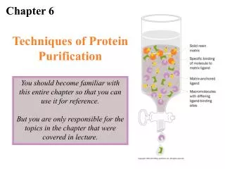
GST pull down assay Principle a simple technique to test interaction between a tagged protein or the bait (GST, His6, biotin ...) and another protein (test protein, or prey). Method
|BamH1 ||EcoR1 ||SmaI |SalI | XhoI | NotI | ... ATC GAA GGT CGT GGG ATC CCC AGG AAT TCC CGG GTC GAC TCG AGC GGC CGC ... ... TAG CTT CCA GCA CCC TAG GGG TCC TTA AGG GCC CAG CTG AGC TCG CCG GCG ... ... Ile Glu Gly Arg Gly Ile Pro Arg Asn Ser Arg Val Asp Ser Ser Gly Arg ... | Factor Xa | 1. Engineered protease site allows removal of fusion partner
GST pull-down assay Sepharose GSH “Y” “Y” GST “X” GST Sepharose GSH
GST pull-down assay Sepharose GSH “Y” GST “X” GST Sepharose GSH
GST pull-down assay Run Western blot Sepharose GSH “Y” Input GST-X GST GST “X” anti-Y
2. Addition of a few residues should have minimal effect on recombinant protein • His6 Tag • add 6 consecutive His to either end • binds metals • Epitope Tag • 6-12 amino acids • mAb for detection or purification
Immunoprecipitation • affinity purification based on isolation of Ag-Ab complexes • analyze by gel electrophoresis • initially based on centrifugation of large supramolecular complexes • [high] and equal amounts • isolation of Ag-Ab complexes • fixed S. aureus • protein A-agarose • protein G-agarose • Bacterial proteins that bind IgG (Fc): • protein A (Staphylococcus aureus) • protein G (Streptococcus) • binds more species and subclasses
Immunoprecipitation(면역침강) Method Cell lysate 준비 Cell lysate Preclearing Immunoprecipitation과정 시 방해가 될 수 있는 chromosome이나 Proteins A or Proteins G에 친화성이 강해 antigen-antibody의 침전과정과 별개로 직접 the bead에 붙을 수 있는 것들을 없애주기 위함 Immunoprecipitation precleared lysate가 들어있는 tube에 1~10ug의 antibody을 넣어줍니다 Washing Ab와 결합하지 않은 상층액을 제거후 Washing buffer를 이용하여 washing 하여 줌 SDS-PAGE loading 후 wastern blotting 으로 확인 DNase foot printing method Figure. Immunoprecipitation
agarose Typical IP Protocol • 1. Solubilize antigen • usually non-denaturing • SDS + excess of TX100 • 2. Mix extract and Ab • 3. Add protein G-agarose, etc • 4. Extensively wash • 5. Elute with sample buffer • 6. SDS-PAGE • 7. Detection • protein stain • radioactivity G
Rb inactive E2F ATP p p active Rb + E2F gene expression DNA replication Example : MIC-1 (ng/ml) 0 20 50 Rb IP : Rb E2F CD9 co-immunoprecipitates with aIIb3 in Brij-35- (BRIJ), but not TX-100-solubilized platelets Detergent Strong : TritonX-100, NP-40 Mild : Brij, CHAPS
partner target Flag, Myc, HA, GFP… antibody Fusion Proteins • increase stability • affinity purification • detection/assay • spectrophotometric • binding assays • antibodies • export signals
두가지 type의 hybrid를 만듬 DBD (DNA binding domain) – protein AD (activation domain) – protein : DBD와 AD가 동시에 존재할 경우만이 유전자의 promoter region에 binding 하여 유전자의 발현을 촉진 시킴 만들어진 두 가지 type의 protein을 세포 내에 도입시키고 유전자의 발현을 관찰 주로 reporter gene 을 통하여 간접적으 로 관찰함 yeast를 이용할 경우는 minimal medium 에서 자랄 수 없던 것이 형질이 전환되어 mm 에서도 자랄 수 있는 것과 같은 특징 이용 함 The yeast two hybrid system Principle protein-protein interactions 을 알아 보기 위한 방법 Eukaryote의 경우 complex를 이루어서 signal을 전달하는 경우가 많은데 이렇듯 complex를 이루어서 작용하는 protein을 찾아 낼 경우 사용하는 방법 protein의 DNA binding domain과 activation domain 분리하여 각각에 binding 할 것이라 생각되는 protein을 붙인 후 yeast의 형질 전환을 통해 protein의 binding을 예측 함 Method Figure. Yeast two hybrid system
AD DBD + gene fish AD bait DBD AD DBD Measurable product + reporter
reporter DBD bait HIS lacZ his- leu- trp- AD fish trp AD DBD leu Measurable product his nucleus Yeast 2-Hybrid Assay
Example Oncogene (2003) 22, 6151 - 6159 : Xiaoying Yin, Christine Giap and John S Lazo
Example Figure. PIAS 와 Smad 간의 물리적인 결합 확인 Purpose PIAS가 Smad6, 7과 binding 하는지 알아 봄 yeast GAL4 DB - Smad7,6 MH2 yeast GAL4 AD - PIYS mm medium 이용 – 두 가지의 결합 확인 가능 Result Smad 7과 PIYS가 함께 도입된 yeast 만이 생존 각각을 도입한 경우 자라지 않음. 즉 두 가지가Binding 함을 알 수 있음 JBC. Vol, 278, pp 34253- 34258 : Swiyu Imoto, Kenji Sugiyama and Ryuta Muromoto
Gene expression이 왜 중요한가 Signal Signal 세포의 성장세포의 특성세포의 변화세포의 죽음 세포의 기능세포의 역할 A cell B cell Behavior Behavior Behavior
DEFINITION Human mRNA for p53 cellular tumor antigen ACCESSION X02469 M60950 SOURCE human ORGANISM Homo sapiens AUTHORS Zakut-Houri,R., Bienz-Tadmor,B., Givol,D. and Oren,M. TITLE Human p53 cellular tumor antigen: cDNA sequence and expression in COS cells JOURNAL EMBO J. 4 (5), 1251-1255 (1985) FEATURES Location/Qualifiers source 1..1317 /organism="Homo sapiens" /db_xref="taxon:9606" CDS 136..1317 /note="p53 tumor antigen (aa 1-?)" /codon_start=1 /protein_id="CAA26306.1" /db_xref="PID:g35210" /db_xref="GI:35210" /db_xref="SWISS-PROT:P04637" /translation="MEEPQSDPSVEPPLSQETFSDLWKLLPENNVLSPLPSQAMDDLM LSPDDIEQWFTEDPGPDEAPRMPEAAPPVAPAPAAPTPAAPAPAPSWPLSSSVPSQKT YQGSYGFRLGFLHSGTAKSVTCTYSPALNKMFCQLAKTCPVQLWVDSTPPPGTRVRAM AIYKQSQHMTEVVRRCPHHERCSDSDGLAPPQHLIRVEGNLRVEYLDDRNTFRHSVVV PYEPPEVGSDCTTIHYNYMCNSSCMGGMNRRPILTIITLEDSSGNLLGRNSFEVRVCA CPGRDRRTEEENLRKKGEPHHELPPGSTKRALPNNTSSSPQPKKKPLDGEYFTLQIRG RERFEMFRELNEALELKDAQAGKEPGGSRAHSSHLKSKKGQSTSRHKKLMFKTEGPDSD"
CDS 136..1317 ORIGIN 1 gtctagagcc accgtccagg gagcaggtag ctgctgggct ccggggacac tttgcgttcg 61 ggctgggagc gtgctttcca cgacggtgac acgcttccct ggattggcag ccagactgcc 121 ttccgggtca ctgccatgga ggagccgcag tcagatccta gcgtcgagcc ccctctgagt 181 caggaaacat tttcagacct atggaaacta cttcctgaaa acaacgttct gtcccccttg 241 ccgtcccaag caatggatga tttgatgctg tccccggacg atattgaaca atggttcact 301 gaagacccag gtccagatga agctcccaga atgccagagg ctgctccccc cgtggcccct 361 gcaccagcag ctcctacacc ggcggcccct gcaccagccc cctcctggcc cctgtcatct 421 tctgtccctt cccagaaaac ctaccagggc agctacggtt tccgtctggg cttcttgcat 481 tctgggacag ccaagtctgt gacttgcacg tactcccctg ccctcaacaa gatgttttgc 541 caactggcca agacctgccc tgtgcagctg tgggttgatt ccacaccccc gcccggcacc 601 cgcgtccgcg ccatggccat ctacaagcag tcacagcaca tgacggaggt tgtgaggcgc 661 tgcccccacc atgagcgctg ctcagatagc gatggtctgg cccctcctca gcatcttatc 721 cgagtggaag gaaatttgcg tgtggagtat ttggatgaca gaaacacttt tcgacatagt 781 gtggtggtgc cctatgagcc gcctgaggtt ggctctgact gtaccaccat ccactacaac 841 tacatgtgta acagttcctg catgggcggc atgaaccgga ggcccatcct caccatcatc 901 acactggaag actccagtgg taatctactg ggacggaaca gctttgaggt gcgtgtttgt 961 gcctgtcctg ggagagaccg gcgcacagag gaagagaatc tccgcaagaa aggggagcct 1021 caccacgagc tgcccccagg gagcactaag cgagcactgc ccaacaacac cagctcctct 1081 ccccagccaa agaagaaacc actggatgga gaatatttca cccttcagat ccgtgggcgt 1141 gagcgcttcg agatgttccg agagctgaat gaggccttgg aactcaagga tgcccaggct 1201 gggaaggagc caggggggag cagggctcac tccagccacc tgaagtccaa aaagggtcag 1261 tctacctccc gccataaaaa actcatgttc aagacagaag ggcctgactc agactga
ATG? ATG? 5’ end of 1st exon missing when cDNA cloning can be determined by 1) Primer extension 2) RNase protection assay (RNase mapping) 3) S1 nuclease assay 4) 5’-RACE (Rapid Amplification of cDNA End)
Trancription machinery components General (Basal) factors Upstream factors ubiquitous not regulated initiation efficiency Inducible factors similar to upstream factors regulatory role at specific time and specific tissue binding site is called "response element" Definition of promoter and enhancer Promoter : responsible for only initiation (~200 bp) Enhancer : enhance initiation, closely packed array (~100 bp)
Module Consensus Factor Comments TATA box TATAAAA TBP -25 ; initiation precision CAAT box GGCCAATCT CTF/NF1 CP1, 2, 3, 4 C/EBP, ACF -75 ; either orientation GC box GGGCGG Sp1 -90 ; either orientation, often multicopy Sp1 is a monomer, 105 kDa Octamer ATTTGCAT Oct-1 Oct-2 Oct-1 is ubiquitous Oct-2 is lymphoid specific factor B GGGACTTTCC NFB ATF GTGACGT ATF Promoter Characterization It is not possible to predict the DNA sequence recognized by proteins 1) Upstream factors Basal factor (TATA, Inr) : initiation location Upstream factor (GC, CAAT) frequency of initiation (assembly) Conserved element does not inevitably imply binding of protein
Module Consensus Factor Regulatory agent HSE CNNGAANNTCCNNG HSTF heat shock GRE TGGTACAAATGTTCT Receptor glucocorticoid TRE TGACTCA AP1 phorbol ester SRE CCATATTAGG SRF serum 2) Response elements May be located in promoters or enhancers Active protein is available only under certain condition Any one of several elements can independently activate the gene
3) Character of transcription factors Structure ① DNA-binding domain (usually basic) ② Activation domain (usually acidic) ③ Dimerization domain Domains are independent, interchangable
Transactivation direct interaction or with coactivators contact with TFIID (most common, esp TAFs), TFIIB, TFIIA can influence to initiation complex by looping at a distance
Categories of Transcription Activators according to DBD, [Gene VII] 1) Helix-turn-helix (HTH) motif 2) Zn finger motif3) Leucine zipper (Zip) motif, usually basic (bZip) 4) Helix-loop-helix (HLH) motif, usually basic (bHLH) 1) Helix-Turn-Helix motif two or three helices and short chain (turn) can form dimer Homeodomain proteins found in proteins related to development originally found in Drosophila determine the identity of body structure also found in higher eukaryote Oct proteins ; 75 aa, called Pou domain
2) Zinc Finger motif helix - Zn - sheet helix contacts DNA Classic zinc finger proteins : Sp1 (3 fingers) Steroid receptors : 2 fingers steroid hormone : MR, AR, PR, ER thyroid hormone : T3R retinoic acid (vitamin A) : RAR, RXR bind to a specific receptor that activates gene transcription "receptor" may be a misnomer recognize special consensus sequence, like GRE consist of central DNA binding domain N-terminal activation domain C-terminal ligand binding domain
Additional) b-barrels two sheets contacts DNA Papilloma virus activator E2 3) Leucine Zipper motif two helices DNA binding and protein dimerization by same motif homodimer or heterodimer expand the repertoire of DNA-binding specificities basic region is DNA binding domain, bZip C/EBP Jun/Fos, JunB, JunD, Fra (Fos-related Antigen) Fos cannot homodimerize Jun/Fos can bind with an activity more 10 folds than Jun/Jun
4) Helix-Loop-Helix motif short helix and long helix homodimer or heterodimer highly basic region can bind to DNA ; bHLH E12, E47 (Ig gene enhancer) MyoD, myogenin, Myf-5 (myogenesis), Myc (oncogene) bHLH fall into 2 groups class A : ubiquitously expressed (E12/E47) class B : tissue-specific manner (MyoD) some HLH protein lacks long helix can dimerize, unable to bind dominant negative way (Id proteins)
PROMOTER ANALYSIS 1) Oocyte system 2) Transfection system CAT assay Luciferase assay 3) Transgenic system 4) in vitro system EMSA (electrophoretic mobility shift assay) DNase I footprinting assay In vitro transcription assay 5) Transcription factor characterization Affinity chromatography Two hybrid 6) in vivo system in vivo DNase I footprinting assay ChIP assay (chromatin immunoprecipitation)
RAR RXR reporter Measurable product nucleus How to measure gene activation in eukaryotic cells ? Transfection assays
RAR Hormone (steroid) RXR reporter Measurable product nucleus How to measure gene activation in eukaryotic cells ? Transfection assays