Download
1 / 7
70 likes | 167 Views
This study explores how nuclear substrates like Mat2 are exported to the cytoplasm through Doa10, investigating INM localization and targeted silencing. Ultrastructural data and targeted degradation assays provide insights into Doa10's role.

E N D
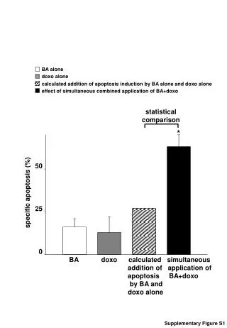
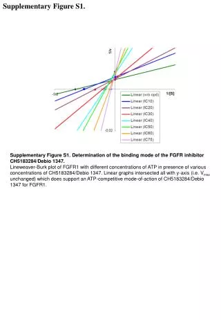
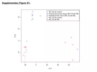
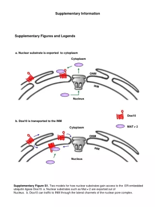
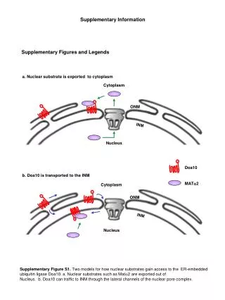
a. Nuclear substrate is exported to cytoplasm Cytoplasm ONM INM Nucleus Doa10 b. Doa10 is transported to the INM MAT2 Cytoplasm ONM INM Nucleus Supplementary Figure S1. Two models for how nuclear substrates gain access to the ER-embedded ubiquitin ligase Doa10. a. Nuclear substrates such as Mat2 are exported out of Nucleus. b. Doa10 can traffic to INM through the lateral channels of the nuclear pore complex. Supplementary Information Supplementary Figures and Legends
NE N 0.1 m 0. 06 m Supplementary Figure S2. Ultrastructural localization of Doa10 to the inner NE. Anti-GFP immunogold EM staining was used to localize Doa10-GFP. Arrowheads mark gold beads. Left: Cell without Nup53 overexpression. Right: Nup53-induced INM lamellae.
His – His – Trp – GBD Aeb:UASG GBD-Stt3 GBD-Doa10 aeB GBD-Doa10 aeb:UASG GBD-Doa10 Aeb:UASG sir2 GBD-Doa10 Supplementary Figure S3. Targeted silencing by GBD-Doa10 assayed with serially diluted yeast cells. Proteins were expressed in YSB35 (top three rows); YSB1 (no UASG); YSB41, which has UASG but lacks all three HMR-E elements; and RS1132, a sir2 mutant7 .
HMG1HC nup53∆C 2m Sec61-GFP Supplementary Figure S4. Overexpression of Hmg1 and Nup53Cfail to induce theta nuclei. Sec61-GFP-expressing cells were transformed with either a high-copy HMG1 or nup53C plasmid and imaged by confocal microscopy.
100 80 60 gal activity (%) doa10∆ 40 pom152∆ 20 nup188∆ WT 0 0 15 30 60 90 Chase time (min) Supplementary Figure S5. Partial impairment of Doa10 import in nup188 and pom152mutants correlates with a mild defect in the degradation of a nuclear substrate, Deg1-bgal. Degradation was measured by bgal activity assays of three independent cultures after addition of cycloheximide. Half-lives were significantly longer in nup188 (P<0.01) and pom152 (P<0.05) relative to WT.
Doa10 Crn1 ER N Cyt Supplementary Figure S6. Scheme to tether Doa10 at cortical ER sites and prevent INM entry. The actin-binding domain of Crn1, which binds to cortical actin patches, was fused to Doa10 (followed by GFP), yielding Doa10-CNG.
100 80 doa10 doa10- pgk1 60 gal activity (%) 40 WT 20 doa10- hrd1C 0 0 30 60 90 Chase time (min) doa10- pgk1 doa10- hrd1C doa10∆ WT 15 30 0 15 30 0 15 30 0 0 15 30 Ura3-SL17 Pgk1 a b Supplementary Figure S7. a. Nuclear substrate degradation correlates with Doa10 nuclear entry. Deg1-bgal degradation was measured as in Suppl.Fig. 5.b. Cytoplasmic substrate turnover is not impaired by Doa10 nuclear exclusion.