Download
1 / 38
600 likes | 1.55k Views
Acute Exacerbation of Chronic Obstructive Pulmonary Disease. Prof. Ashraf M. Hatem, MD, FCCP. Definition of Acute exacerbation:.

E N D
Acute Exacerbation of Chronic Obstructive Pulmonary Disease. Prof. Ashraf M. Hatem, MD, FCCP
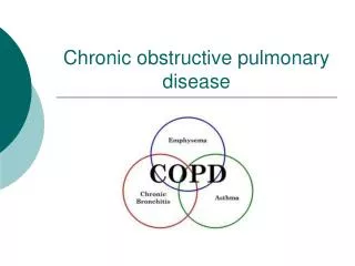
Definition of Acute exacerbation: • The definition of COPD exacerbation is an acute change in a patient’s baseline dyspnoea, cough and/or sputum beyond day-to-day variability sufficient to warrant a change in therapy. • Causes of exacerbation can be both infectious and non-infectious e.g. air pollution.
Most commonly encountered organisms: -Streptococcus pneumoniae -Hemophilus influenzae -Moraxella catarrhalis • The cause in one third of exacerbations remains unidentified
Classification of Severity of Acute Exacerbation of COPD • The Operational Classification of Severity is as follows: • Level I: ambulatory (outpatient), • Level II: requiring hospitalisation, and • Level III: acute respiratory failure.
The Operational Classification of Severity of COPD exacerbation
Indications for hospitalisation of patients with a COPD exacerbation • Presence of high-risk co-morbid conditions, including pneumonia, cardiac arrhythmia, congestive heart failure, diabetes mellitus, renal or liver failure • Inadequate response of symptoms to outpatient management • Marked increase in dyspnoea • Inability to eat or sleep due to symptoms • Worsening hypoxaemia • Worsening hypercapnia • Changes in mental status • Inability of the patient to care for her/himself • Uncertain diagnosis • Inadequate home care
Level III: treatment in patients requiring special or intensive care unit
In-patient Oxygen Therapy • The goal is to prevent tissue hypoxia by maintaining arterial oxygen saturation (Sa,O2) at >90%. • Main delivery devices include nasal cannula and venturi mask. • Alternative delivery devices include nonrebreather mask, reservoir cannula, nasal cannula or transtracheal catheter.
Arterial blood gases should be monitored for arterial oxygen tension (Pa,O2), arterial carbon dioxide tension (Pa,CO2) and pH. • Arterial oxygen saturation as measured by pulse oximetry (Sp,O2) should be monitored for trending and adjusting oxygen settings.
Prevention of tissue hypoxia supersedes CO2 retention concerns. • If CO2 retention occurs, monitor for acidosis. • If acidaemia occurs, consider mechanical ventilation.
MEASURES TO MOBILIZE AIRWAY SECRETIONSIN HOSPITALIZED PATIENTS WITH COPD • Directed coughing, “huff coughing.” Benefit extrapolated from experience in cystic fibrosis • Chest physiotherapy: manual or mechanical chest percussion and postural drainage. Benefit extrapolated from experience in cystic fibrosis. Can cause transient fall in FEVI. Assumed role limited to patients with > 25 ml sputum per day or lobar atelectasis from mucus plugging • Intermittent positive pressure breathing (IPPB). Not indicated; no proven benefit In COPD • Positive expiratory pressure (PEP). Benefit extrapolated from experience in cystic fibrosis. No reported experience in acute exacerbations of COPD.
Bland aerosol therapy. No demonstrated benefit in COPD unless artificial airway is in place. May cause bronchospasm in nonintubated patients. • Systemic hydration. No demonstrated benefit beyond repletion of intravascular volume to euvolemia. • Nasotracheal suctioning. Limited benefit; tolerated only for short periods • Mini-tracheotomy. Possible temporary benefit in patients with persistent airway secretions causing respiratory deterioration.
Indications for ICU Admission • Severe dyspnea that responds inadequately to initial emergency therapy. • Confusion, lethargy, coma. • Persistent or worsening hypoxemia (PaO2 < 5.3 kPa, 40 mm Hg), and/or severe/worsening hypercapnia • (PaCO2 > 8.0 kPa, 60 mm Hg), and/or severe/worsening respiratory acidosis (pH < 7.25) despite supplemental • oxygen and NIPPV.
Assisted ventilation • Noninvasive positive pressure ventilation (NPPV) should be offered to patients with exacerbations when, after optimal medical therapy and oxygenation, respiratory acidosis (pH <7.36) and or excessive breathlessness persist. All patients considered for mechanical ventilation should have arterial blood gases measured.
If pH <7.30, NPPV should be delivered under controlled environments such as intermediate intensive care units (ICUs) and/or high-dependency units. • If pH <7.30, NPPV should be delivered under controlled environments such as intermediate intensive care units (ICUs) and/or high-dependency units.
If pH <7.25, NPPV should be administered in the ICU and intubation should be readily available. • The combination of some continuous positive airway pressure (CPAP) (e.g. 4–8 cmH2O) and pressure support ventilation (PSV) (e.g. 10–15 cmH2O) provides the most effective mode of NPPV. • Patients meeting exclusion criteria should be considered for immediate intubation and ICU admission.
Exclusion criteria include: • respiratory arrest, • cardiovascular instability, • impaired mental status, • somnolence, • inability to cooperate, • copious and/or viscous secretions with high aspiration risk, • recent facial or gastro-oesophageal surgery; craniofacial trauma and/or fixed naso-pharyngeal abnormality, • burns, • extreme obesity. • In the first hours, NPPV requires the same level of assistance as conventional mechanical ventilation.
Indications for Mechanical Ventilation • Severe dyspnea with use of accessory muscles an paradoxical abdominal motion. • Respiratory frequency > 35 breaths per minute. • Life-threatening hypoxemia (PaO2 < 5.3 kPa, 40 mm Hg or PaO2/FiO2 < 200 mm Hg). • Severe acidosis (pH < 7.25) and hypercapnia (PaCO2 > 8.0 kPa, 60 mm Hg).
Respiratory arrest. • Somnolence, impaired mental status. • Cardiovascular complications (hypotension, shock, heart failure). • Other complications (metabolic abnormalities, sepsis, pneumonia, pulmonary embolism, barotrauma, massive pleural effusion). • NIPPV failure (or contraindication to NIPPV).
Mechanical Ventilation • Assisted ventilation should be considered for patients with acute exacerbations of COPD when pharmacologic and other nonventilatory treatments fail to reverse clinically significant respiratory failure. • The clinician must aim to avoid complications associated with mechanical ventilation and should initiate weaning and discontinuation of mechanical ventilation as soon as possible.
The main goals of assisted positive pressure ventilation in acute respiratory failure complicating COPD are: - Resting of ventilatory muscles, and - Restoration of gas exchange to a stable baseline. • Allow for permissive hypercapnea (except in cerebral edema, myocardial ischemia, LVF….)
There are three specific pitfalls in ventilating patients with COPD: i- Overventilation, resulting in acute respiratory alkalemia, ii- Initiation of complex pulmonary and cardiovascular interactions that may result in systemic ypotension. iii- Creation of intrinsic positive end-expiratory pressure (PEEP), or “auto-PEEP,” especially if expiratory time is inadequate or if dynamic airflow obstruction exists
The three ventilatory modes most widely used for managing patients with COPD are: - Assist-control ventilation (ACV), - Intermittent mandatory ventilation (IMV), and - Pressure support ventilation (PSV). • PSV provides increased patient comfort, promotes patient synchrony with the ventilator, and facilitate weaning from mechanical ventilation in the patient who maintains adequate ventilatory drive.
GOLD Guidelines: Treatment of COPD
Discharge Criteria for Patients With Exacerbations of COPD • Inhaled ß2-agonist therapy is required no more frequently than every 4 hrs. • Patient, if previously ambulatory, is able to walk across room. • Patient is able to eat and sleep without frequent awakening by dyspnea. • Patient has been clinically stable for 12-24 hrs.
Arterial blood gases have been stable for 12-24 hrs. • Patient (or home caregiver) fully understands correct use of medications. • Follow-up and home care arrangements have been completed (e.g., visiting nurse, oxygen delivery, meal provisions). • Patient, family, and physician are confident patient can manage successfully.
Strategies to Help the PatientWilling to Quit Smoking (5 As) • ASK: Systematically identify all tobacco users at every visit. Implement an office-wide system that ensures that, for EVERY patient at EVERY clinic visit, tobacco-use status is queried and documented. • ADVISE: Strongly urge all tobacco users to quit. In a clear, strong, and personalized manner, urge every tobacco user to quit. • ASSESS: Determine willingness to make a quit attempt. Ask every tobacco user if he or she is willing to make a quit attempt at this time (e.g., within the next 30 days). • ASSIST: Aid the patient in quitting. Help the patient with a quit plan; provide practical counseling; provide intra-treatment social support; help the patient obtain extra-treatment social support; recommend use of approved pharmacotherapy except in special circumstances; provide supplementary materials. • ARRANGE: Schedule follow-up contact. Schedule follow-up contact, either in person or via telephone.
Long Term Oxygen Therapy • Oxygen administration >15 hours/day. • Indications: - PaO2< 55 mmHg or SaO2< 88% - PaO2 55-60 mmHg or SaO2 > 89% if there is evidence of Pulmonary hypertension, CHF, or Polythycemia (Hematocrit > 55%)
Pulmonary Rehabilitation • Goals: - Reduce symptoms - Improve QOL - Promote physical and emotional participation in every day life • Program should be at least 2 months. • Aim is to strengthen inspiratory muscles and increase endurance.
Surgical Treatment • Bullectomy • LVRS and Bronchoscopic volume reduction • Lung transplantation
Assessment 4-6 Weeks After Discharge from Hospital • Ability to cope in usual environment. • Measurement of FEV1. • Reassessment of inhaler technique. • Understanding of recommended treatment regimen. • Need for long-term oxygen therapy and/or home • Nebulizer (for patients with very severe COPD).