Download
1 / 32
320 likes | 439 Views
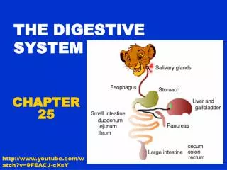
The human digestive system, also known as the alimentary canal, consists of a continuous tube connecting the mouth to the anus, featuring two openings for the intake and expulsion of food. This system includes organs such as the mouth, pharynx, esophagus, stomach, small intestine, and large intestine, each playing crucial roles in digestion. Accessory organs, including the pancreas, liver, and gallbladder, aid in the process. This overview will explore the anatomy and physiological functions, highlighting key components like tunics, sphincters, and surface structures that enhance digestion.

E N D
The Digestive System The Anatomy
Anatomy • We have what we call an alimentary canal • This means we have 2 holes • One for import, one for export • This is in contrast to organisms such as jellyfish who have a gastrovascular cavity • You guessed it…..just one hole for both in AND out • Moral of the story: be glad you have 2 holes!!
Anatomy • Connecting our 2 holes we have a series of tubes PLUS a bunch of accessory organs that assist in the digestion of foods
The Tubes • Our alimentary canal, aka our gastrointestinal (GI) tract, consists of a continuous, coiled, hollow, muscular tube • Organs • Mouth • Pharynx • Esophagus • Stomach • Small intestine • Large intestine
1) Mouth • Lined with a mucous membrane • Question: What type of membrane is that? • Epithelial membrane • Means it is comprised of epithelial tissue and connective tissue • Important components • Tongue • Teeth • Salivary glands
2) Pharynx • Passageway for food, fluids, and air • Muscular passageway to propel food • Called peristalsis
CAREFUL!! • Do NOT confuse this structure with the larynx • The pharynx is for phood (food) and air • The larynx is for voice (we will talk about this in the respiratory system) • Food should NOT be found here!!
True or False Food passes through the larynx on the way to the stomach.
3) Esophagus • Runs from the pharynx to the stomach • 4 layers (innermost to outermost) called tunics • Mucosa • Moist membrane that lines the lumen (the hollow part of the tube) • Epithelial tissue, connective tissue, smooth muscle layer • Submucosa • Connective tissue • Blood vessels, nerve endings, lymph organs
3) Esophagus • Muscularisexterna • Inner circular layer and outer longitudinal layer of smooth muscle • Serosa • Outermost layer consisting of flat serous fluid-producing cells • Visceral peritoneum and parietal peritoneum
Question • What are the 4 tunics, in order, from external to internal? • Serosa, muscularisexterna, submucosa, mucosa
Location Matters • Notice the location of the esophagus to the trachea
Question • What prevents our food from “going down the wrong tube”? • Answer: the epiglottis!! • Flap of cartilage that directs food to the stomach and air to the lungs
4) Stomach • C-shaped organ • Has the same 4 tunics as the esophagus • What were those again? • Mucosa, submucosa, muscularisexterna, serosa
4) Stomach • Has 2 sphincters • These acts as gatekeepers letting things in and out of the stomach • Cardioesophageal sphincter connects the cardiac region of the stomach (closest to the heart) with the esophagus • the pyloric sphincter connects the stomach to the small intestine • Let’s add to our picture!
4) Stomach • Contains large folds of mucosa called rugae • This enables it to be flexible in its diameter • When full, it can hold up to ONE GALLON of food
Surgery: Gastric Bypass • Treatment for obesity • Involves decreasing the size of the available storage space of the stomach • Intended to make you feel full faster so that way you can eat less • Surgery Squad! • http://www.surgerysquad.com/surgeries/rny-gastric-bypass-surgery/
4) Stomach: Microscopic • Pitted appearance called gastric pits that lead to gastric glands • There is also a variety of cells that assist in digestion • (We will get to those when we get to the physiology of digestion )
5) Small Intestine • Connects to the stomach via the pyloric sphincter • Coiled tube • Average length: 8-18 feet!! • Same 4 tunics of the stomach and esophagus • 3 subdivisons • Duodenum (5% of length) • Jejunum (40% of length) • Ilium (55% of length)
5) Small Intestine • Contains ducts that connect to accessory organs • Both ducts merge into 1 duct • Pancreatic duct • Connects the pancreas to the small intestine • Common bile duct • Connects the liver/gall bladder to the small intestine • The gall bladder, liver, and pancreas are accessory organs to the GI tract
5) Small Intestine: Microscopic • The small intestine has folds in it, just like the stomach • Called circular folds • Difference: these folds are not for stretching like the rugae of the stomach • These folds are for increased surface area
5) SI: Microscopic • More microscopically, villi are fingerlike projections of mucosa found facing the lumen of the SI on the circular folds • Again, these function to increase the surface area
5) SI: Microscopic • Even more microscopically, there are a series of microvilli on each villus • Projections of the plasma membrane of the mucosa cells • Often referred to as the “brush border”
Let’s Draw a Picture • Circular folds • Villi • Microvilli
Question • Which of the following has the 4 tunics? • A) esophagus • B) SI • C) stomach • D) LI • E) all except D
6) Large Intestine • Larger in diameter than the SI, but shorter in length • Frames the SI on 3 sides • Subdivisions • Cecum attaches the SI to the LI • The appendix hangs off of this structure • Called vermiform appendix because it looks like a rat tail • Colon • Ascending colon • Transverse colon • Descending colon • Sigmoid colon • Rectum • Anal canal
Interesting….. • Your anus is a sphincter • That is, it has a muscular gatekeeper • It has both voluntary and involuntary muscle so technically you can’t always control your elimination • Elimination=defecating=going #2
Quiz Yourself • Put the following organs in order: pharynx, mouth, small intestine, large intestine, stomach, esophagus • Mouth, pharynx, esophagus, stomach, SI, LI • Where does food pass through first: the cardioesophageal sphincter or the pyloric sphincter? • Cardioesophageal and then pyloric • What are the three accessory organs and how are they connected to our GI tract? • Liver, gall bladder, and pancreas; SI • What allows our stomach to expand? • Rugae folds • What is the purpose of the circular folds, villi, and microvilli? What organ contains these structures? • Increase surface area; SI